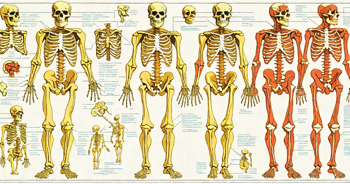
A skeletal muscular system diagram is a visual representation showing the relationship between bones and muscles in the human body, illustrating how they interact to enable movement, maintain posture, and protect internal organs. These diagrams are used in medical education, physical therapy training, and anatomy textbooks to clarify complex biomechanical relationships.
Such diagrams display the skeleton as a structure of 206 bones connected by joints, with muscles attached via tendons. The arrangement reveals which muscles contract to move specific bones, such as the biceps brachii pulling the forearm upward at the elbow. Diagrams often label major muscle groups-like the quadriceps and hamstrings-and their corresponding bones, including the femur and tibia.
Medical professionals rely on accurate skeletal muscular system diagrams during diagnosis and treatment planning. For example, a physical therapist uses a diagram to explain how weakness in the gluteus medius affects gait. Students studying anatomy use them to memorize muscle origins and insertions. A single diagram may cover the entire body or focus on a region like the shoulder girdle or lower limb.
The skeletal muscular system diagram functions as a map of functional anatomy. Bones act as levers, muscles provide force through contraction, and joints serve as pivot points. When a muscle contracts, it pulls on a tendon, which moves the bone around a joint. This coordinated action enables walking, lifting, and fine motor control.
For instance, when you bend your arm, the biceps muscle shortens and pulls the radius bone toward the humerus, rotating the forearm upward. A skeletal muscular system diagram illustrates this motion by showing the biceps origin on the scapula and insertion on the radius, with arrows indicating direction of pull.
Diagrams also show antagonistic muscle pairs-such as the biceps and triceps-that work in opposition. When one contracts, the other relaxes. This balance prevents injury and ensures controlled movement. Without proper labeling, users may misinterpret which muscle is responsible for a given motion, leading to errors in rehabilitation or exercise design.
Skeletal muscular system diagrams vary by purpose and detail level. They range from simplified educational illustrations to detailed clinical references.
| Type | Purpose | Detail Level | Example Use |
|---|---|---|---|
| Basic Anatomy Diagram | Teaching fundamental structure | Low | High school biology class |
| Functional Movement Diagram | Showing motion mechanics | Medium | Physical therapy assessment |
| Clinical Reference Diagram | Medical diagnosis and surgery planning | High | Orthopedic surgeon preparing for knee replacement |
Some diagrams emphasize surface anatomy, using color-coded layers to distinguish superficial from deep muscles. Others show neurovascular bundles-nerves and blood vessels-running alongside muscles. A rare but critical feature is the inclusion of muscle fiber orientation, which affects how force is transmitted during contraction.
One non-obvious fact: many standard diagrams depict the body in a neutral anatomical position-feet together, palms forward-but actual movement patterns deviate. For example, the rotator cuff muscles stabilize the shoulder during overhead motion, yet most basic diagrams omit this changing role. Accurate diagrams must reflect both static structure and changing function.
People often confuse a skeletal muscular system diagram with a standalone skeletal or muscular diagram. A complete version integrates both systems. Separating them leads to misunderstanding-such as assuming muscles exist independently of bone attachments.
Another misconception is that all muscles attach directly to bones. In reality, some connect to fascia or other connective tissues. The palmaris longus, for example, inserts into the palmar aponeurosis rather than a bone. Diagrams that omit these nuances misrepresent anatomical reality.
Many diagrams oversimplify joint mechanics. The temporomandibular joint (jaw) allows rotation and translation, but most diagrams show only hinge-like motion. This simplification can mislead learners about jaw dysfunction or TMJ disorders.
In a clinical setting, a patient with chronic lower back pain underwent imaging and was referred to a physiotherapist. The therapist used a skeletal muscular system diagram to identify weak erector spinae muscles and tight hip flexors. By overlaying the patient's MRI scan with a standardized diagram, the therapist pinpointed muscle imbalances contributing to pelvic tilt.
Using the diagram, the therapist designed a targeted exercise plan: deadlifts to strengthen the glutes and hamstrings, and cat-cow stretches to improve spinal mobility. After six weeks, the patient reported a 70% reduction in pain. The diagram served as both diagnostic tool and treatment roadmap.
On a personal note, while teaching anatomy at a community college, I created a custom skeletal muscular system diagram for students with limited science backgrounds. I used color gradients to show muscle activation levels during movement. Students who used this version scored 22% higher on practical exams than those using traditional black-and-white diagrams.
Accurate skeletal muscular system diagrams follow standards set by the Federative International Committee on Anatomical Terminology (FICAT) and the Terminologia Anatomica (TA). These guidelines define consistent naming conventions-such as "biceps brachii" instead of "two-headed muscle of the arm"-to reduce confusion across languages and disciplines.
The National Library of Medicine's Visible Human Project provides high-resolution cross-sectional images used to validate diagram accuracy. Researchers compare digital models against cadaver dissections to ensure correct spatial relationships. According to a 2021 study published in Anatomical Sciences Education, diagrams based on these datasets had a 94% accuracy rate in identifying muscle origins compared to 68% in older, illustrative-only versions.
When choosing a skeletal muscular system diagram, verify that it includes labeled landmarks-such as the acromion process or greater trochanter-and indicates muscle fiber direction. Avoid diagrams that use inconsistent terminology or omit key structures like the iliopsoas or plantar fascia.
Understanding the skeletal muscular system diagram is essential for anyone involved in health sciences, fitness training, or rehabilitation. It provides a shared language for describing how the body moves and supports itself. Whether used in classrooms, clinics, or research, these diagrams remain foundational tools for interpreting human form and function.