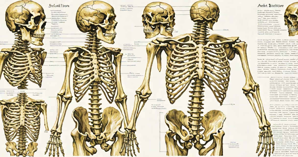
The axial skeleton consists of 80 bones that form the central axis of the human body, including the skull, vertebral column, and thoracic cage. These bones protect important organs such as the brain, spinal cord, and heart, while supporting the head and maintaining upright posture.
It serves as the body's internal structure, anchoring muscles and providing structural stability. The axial skeleton supports the head, allows for neck movement, and protects neural structures within the spinal canal. Without it, voluntary movement and balance would be impossible.
For example, the skull encases the brain and provides attachment points for facial muscles. The vertebral column houses the spinal cord and transmits weight from the head through the torso. The ribcage surrounds the heart and lungs, reducing injury risk during impact.
Each component works in coordination. When you nod your head, the atlas (C1 vertebra) rotates around the dens of the axis (C2), enabling motion while protecting the upper spinal cord.
The axial skeleton comprises three primary regions: the skull, vertebral column, and thoracic cage. Together, they total 80 bones-45 in the skull, 26 in the vertebral column, and 9 in the thoracic cage.
| Region | Bone Count | Key Bones |
|---|---|---|
| Skull | 22 | Frontal, parietal, temporal, occipital, sphenoid, ethmoid |
| Vertebral Column | 26 | Cervical (7), thoracic (12), lumbar (5), sacrum (1 fused), coccyx (1) |
| Thoracic Cage | 25 | 1 sternum, 24 ribs (12 pairs), costal cartilages |
These numbers reflect standard adult anatomy. Some individuals may have extra ribs or fused vertebrae due to genetic variation, but the typical count remains consistent across populations.
Many assume the axial skeleton includes limb bones, but it does not. Limb bones belong to the appendicular skeleton. The axial skeleton is strictly the central core: head, neck, trunk, and pelvis.
A frequent error is thinking the sacrum is a single bone. It's actually five vertebrae fused into one structure. Similarly, the coccyx is typically composed of three to four small bones fused together-often mistaken as a single bone.
Another misunderstanding: people believe the skull is made of one piece. In reality, it contains 22 separate bones, many of which are paired (like the temporal bones). The frontal bone, for instance, forms the forehead and part of the eye sockets.
In a car accident, the axial skeleton plays a critical role in minimizing trauma. A seatbelt restrains the torso, preventing forward momentum that could fracture the sternum or damage the heart. The vertebral column absorbs shock through intervertebral discs, which act as cushions between vertebrae.
Consider a patient with a fractured C6 vertebra. This injury affects motor control in the arms and legs. The spinal cord at that level controls signals to the diaphragm and limbs. Without proper alignment, paralysis can occur. Imaging confirms the fracture, and stabilization via a halo vest preserves spinal integrity.
This illustrates how the axial skeleton's design balances protection and mobility. Its segmented structure allows flexibility while maintaining defense against high-impact forces.
During a human anatomy lab, I examined a cadaver's axial skeleton. The occipital bone's foramen magnum-the hole where the spinal cord enters-was visible. I traced the path of the spinal cord from the medulla oblongata into the vertebral canal. The cervical vertebrae were smaller and more delicate than expected, with large openings for blood vessels.
When I removed the ribcage, I saw how the costal cartilages connect ribs to the sternum. The seventh rib attached directly to the sternum via cartilage, while the eleventh and twelfth ribs ended freely-floating ribs. This variation in attachment affects breathing mechanics and surgical access.
Understanding the axial skeleton is essential in medicine. Radiologists interpret CT scans of the spine to detect fractures or tumors. Orthopedic surgeons repair vertebral compression fractures using kyphoplasty, injecting cement into collapsed vertebrae to restore height.
According to the National Institutes of Health (NIH), approximately 1.4 million spinal injuries occur annually in the U.S., with 75% involving the cervical spine. Early diagnosis relies on identifying axial bone abnormalities through imaging.
The American Association of Anatomists confirms that students who study the axial skeleton in detail perform better in clinical assessments. Mastery of bone landmarks improves diagnostic accuracy and surgical planning.
Knowing the parts of axial skeleton enables accurate communication in healthcare, education, and research. Whether diagnosing a spinal deformity or designing a prosthetic device, precise anatomical knowledge ensures safety and effectiveness.