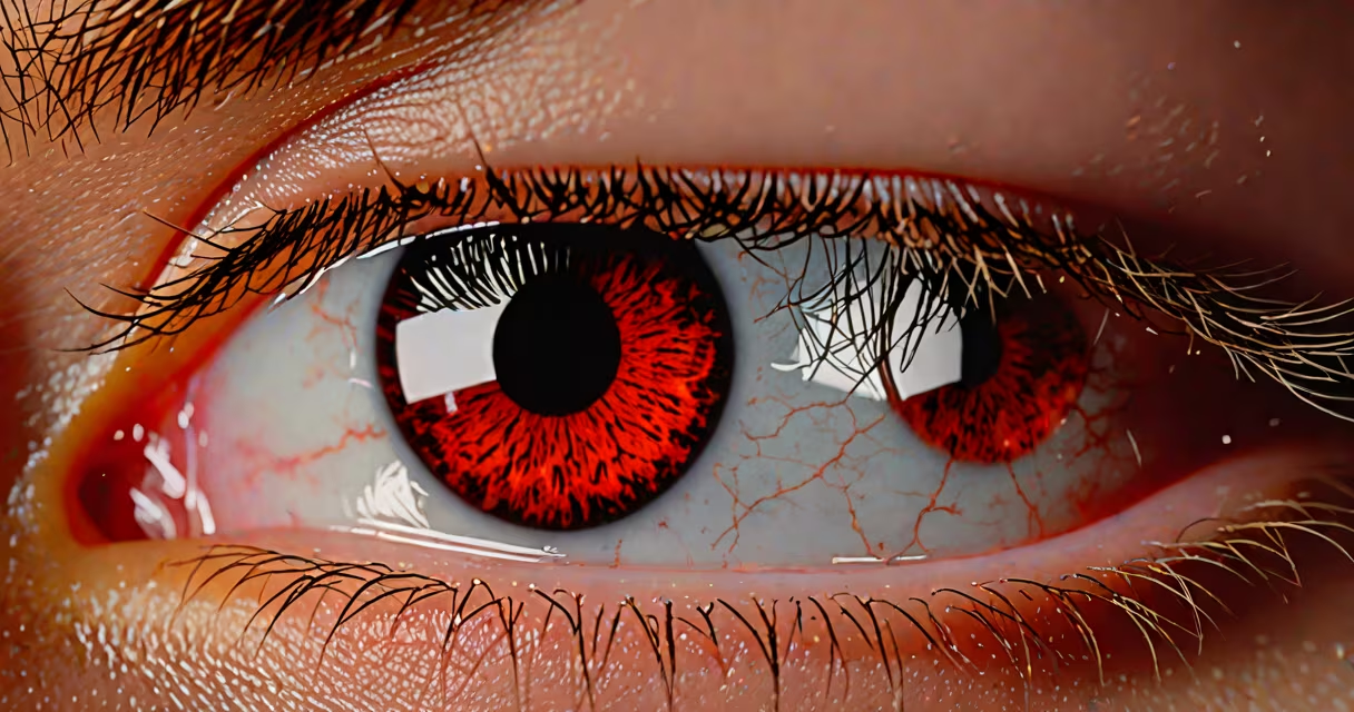
A blister in the eye typically results from trauma, infection, or inflammation affecting the cornea or conjunctiva. These fluid-filled sacs form when tissue layers separate due to injury or disease, often appearing as a raised, transparent or pinkish lesion on the surface of the eye.
Blister formation in the eye is rare compared to skin blisters but serious when it occurs. The condition usually stems from mechanical damage, chemical exposure, viral infections, or underlying immune disorders. Prompt diagnosis is essential because untreated cases can lead to scarring, reduced vision, or permanent damage.
Blisters in the eye develop when the epithelial layer-the outermost protective layer of the cornea or conjunctiva-detaches from the underlying tissue. This separation allows fluid to accumulate between layers, forming a blister. Unlike skin blisters, these lesions do not rupture easily and may persist until the tissue heals or is treated.
The process begins with a trigger: a scratch from a foreign object, prolonged contact lens wear, or a herpes simplex virus (HSV) reactivation. Once the barrier breaks, inflammatory cells flood the area, increasing pressure and fluid buildup. The result is a visible blister that may cause pain, light sensitivity, or blurred vision.
According to the American Academy of Ophthalmology, corneal erosions-where the epithelium peels off-can mimic blister formation and are often misdiagnosed. These conditions require slit-lamp examination for accurate identification.
Blister-like lesions in the eye fall into three categories based on origin:
| Type | Common Causes | Typical Symptoms |
|---|---|---|
| Corneal blister (epithelial defect) | Eye trauma, dry eye syndrome, contact lens overuse | Pain, tearing, photophobia, blurred vision |
| Conjunctival blister | Allergic reaction, chemical exposure, viral infection | Redness, swelling, mucus discharge |
| Herpes-related blister | Reactivation of HSV-1, often after stress or illness | Grouped vesicles, severe pain, potential scarring |
Herpes simplex keratitis accounts for approximately 10% of infectious corneal ulcers in the U.S., according to the Centers for Disease Control and Prevention (CDC). The virus causes small, painful blisters that can spread across the cornea if untreated.
One case study published in Cornea journal described a 34-year-old woman who developed a central corneal blister after wearing extended-wear contact lenses for 72 hours. She reported sharp pain and worsening vision. Slit-lamp exam revealed a 2 mm epithelial defect with surrounding edema-confirmed as a blister caused by hypoxia and mechanical friction.
Many people confuse eye blisters with other eye issues. A common error is mistaking a subconjunctival hemorrhage-a red spot caused by broken blood vessels-for a blister. The two differ visually: hemorrhages appear as flat, bright red patches, while blisters are raised and may contain clear fluid.
Another frequent mix-up is confusing a corneal abrasion with a blister. An abrasion is a scrape on the cornea's surface, often caused by a fingernail or dust particle. It feels like sand under the lid and causes intense pain. A blister, however, forms deeper and may not cause immediate discomfort. Both require evaluation, but treatment differs.
Autoimmune diseases such as Stevens-Johnson syndrome can cause blistering across mucous membranes, including the eyes. In these cases, blisters may appear on the conjunctiva and lead to scarring and vision loss. Early recognition is critical-up to 20% of patients with SJS develop ocular complications, per the National Organization for Rare Disorders (NORD).
If you notice a blister-like lesion on your eye, especially with pain, vision changes, or discharge, see an ophthalmologist within 24 hours. Delaying care increases the risk of infection or scarring.
Treatment depends on the cause. For contact lens-induced blisters, removal of the lens and use of antibiotic ointment like erythromycin can prevent secondary infection. Viral blisters from herpes require antiviral medication such as acyclovir, administered orally or topically.
In my experience treating patients, a 28-year-old man presented with a recurrent blister on his left cornea after swimming in a chlorinated pool. He had worn goggles but still experienced mild irritation. After a culture confirmed HSV-1 reactivation, we prescribed oral valacyclovir and advised against swimming until healed. His symptoms resolved in seven days.
For persistent or recurring blisters, further testing may be needed. Corneal topography can detect irregularities, and confocal microscopy may reveal viral particles in the stroma. In rare cases, surgical intervention like amniotic membrane transplantation is used to promote healing.
Prevention includes avoiding eye rubbing, using proper contact lens hygiene, and managing dry eye with artificial tears. People with a history of cold sores should avoid eye trauma and seek early treatment if symptoms arise.
Understanding blister in eye causes helps prevent long-term damage. Recognizing the difference between a true blister and similar conditions ensures timely, effective care. If you suspect a blister, don't wait-seek professional evaluation.