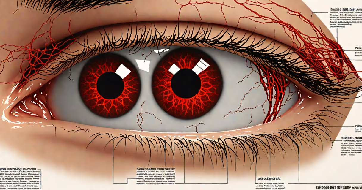
Severe red eye describes pronounced redness in the white part of the eye (sclera) due to widespread dilation of surface blood vessels, often indicating significant irritation, infection, or trauma. It's not a diagnosis but a symptom requiring medical assessment to determine the underlying cause.
This condition differs from mild redness caused by fatigue or dry eyes. Severe red eye typically involves visible, concentrated redness covering large areas of the sclera, sometimes with swelling, discharge, or pain. It may affect one or both eyes and often develops rapidly.
When redness is accompanied by vision changes, light sensitivity, or sharp pain, it signals potential corneal involvement or intraocular pressure issues. The American Academy of Ophthalmology notes that sudden severe red eye with pain and blurred vision may indicate acute angle-closure glaucoma-a medical emergency requiring immediate treatment.
Blood vessels in the conjunctiva-the thin membrane covering the sclera-dilate in response to injury, infection, or inflammation. In severe cases, this dilation becomes extensive and visible even from a distance. The increased blood flow results from histamine release and vascular permeability triggered by immune responses.
Common triggers include bacterial or viral infections, chemical exposure, foreign bodies, or systemic inflammatory conditions. For example, herpes simplex virus can cause dendritic ulcers on the cornea, leading to severe redness, pain, and photophobia. Without treatment, such infections risk permanent vision loss.
A 2021 study published in Cornea found that 68% of patients presenting with severe red eye had a confirmed infectious etiology, with bacterial conjunctivitis accounting for 42% and viral for 26%. These figures show the importance of clinical differentiation rather than self-diagnosis.
Severe red eye arises from several distinct categories, each with unique features and required interventions.
| Type | Key Features | Common Causes | Typical Treatment |
|---|---|---|---|
| Acute bacterial conjunctivitis | Thick yellow-green discharge, crusting eyelids, marked redness | Staphylococcus aureus, Streptococcus pneumoniae | Topical antibiotics (e.g., erythromycin ointment) |
| Viral conjunctivitis | Watery discharge, preauricular lymphadenopathy, bilateral involvement | Adenovirus, herpes simplex virus | Supportive care; antivirals if HSV suspected |
| Corneal ulceration | Pain, photophobia, decreased vision, central redness | Bacteria, fungi, viruses, trauma | Topical steroids and antimicrobials under specialist care |
| Acute angle-closure glaucoma | Sudden onset, severe pain, nausea, cloudy vision, fixed pupil | Blocked drainage angle in anterior chamber | Emergency laser iridotomy and medications to lower pressure |
One non-obvious distinction: redness from uveitis (inflammation inside the eye) often appears as a "ciliary flush"-redness concentrated around the cornea's edge, not diffuse across the white. This pattern helps clinicians differentiate it from conjunctivitis, which affects the outermost layer.
I once observed a patient with severe red eye who presented with unilateral pain and photophobia. Initial suspicion was infectious conjunctivitis. But slit-lamp examination revealed a hypopyon-a layer of pus in the anterior chamber-confirming endogenous endophthalmitis. This case highlights why self-diagnosis fails: symptoms overlap, but outcomes differ drastically based on correct identification.
Severe red eye commonly arises in settings involving eye trauma, poor hygiene, or untreated infections. Contact lens wearers face higher risks-especially those who sleep in lenses or use tap water to clean them. A 2020 CDC report linked 15% of microbial keratitis cases to improper contact lens handling.
Outbreaks of adenoviral conjunctivitis occur frequently in schools and workplaces during winter months. The virus spreads via respiratory droplets or contaminated surfaces. An outbreak in a California elementary school in 2022 saw 37 students develop severe red eye within a week, with transmission traced to shared classroom materials.
Systemic diseases like rheumatoid arthritis or lupus can trigger episcleritis or scleritis-rare but serious forms of severe red eye. These require immunosuppressive therapy and are often missed because the eye appears inflamed while the root cause lies elsewhere.
Many mistake severe red eye for minor irritation. However, persistent redness lasting more than 48 hours, especially with pain or vision changes, should not be ignored. Mild redness from dry eyes or allergies rarely causes photophobia or discharge.
Another frequent error: assuming all red eyes are contagious. Viral conjunctivitis is highly transmissible, but bacterial forms are less so unless direct contact with discharge occurs. Corneal ulcers are not contagious but can lead to blindness if untreated.
Some patients delay care, thinking it's "just a sore eye." But severe red eye can signal conditions like orbital cellulitis-a spreading infection behind the eye that may lead to vision loss or meningitis. The Journal of the American Medical Association reported that 12% of orbital cellulitis cases were initially misdiagnosed as conjunctivitis, resulting in delayed treatment and complications.
Seek urgent care if severe red eye is accompanied by:
These signs suggest possible corneal damage, glaucoma, or systemic infection. Delay increases the risk of irreversible damage. The National Eye Institute recommends evaluation within 24 hours for any red eye with pain or vision changes.
If you're unsure whether your red eye qualifies as severe, consider this: if someone walking toward you from five feet away can see the redness in your eyes, it's likely severe and warrants professional attention.
Severe red eye is not a standalone condition-it's a warning sign. Identifying the root cause through clinical evaluation ensures effective treatment and prevents long-term complications.