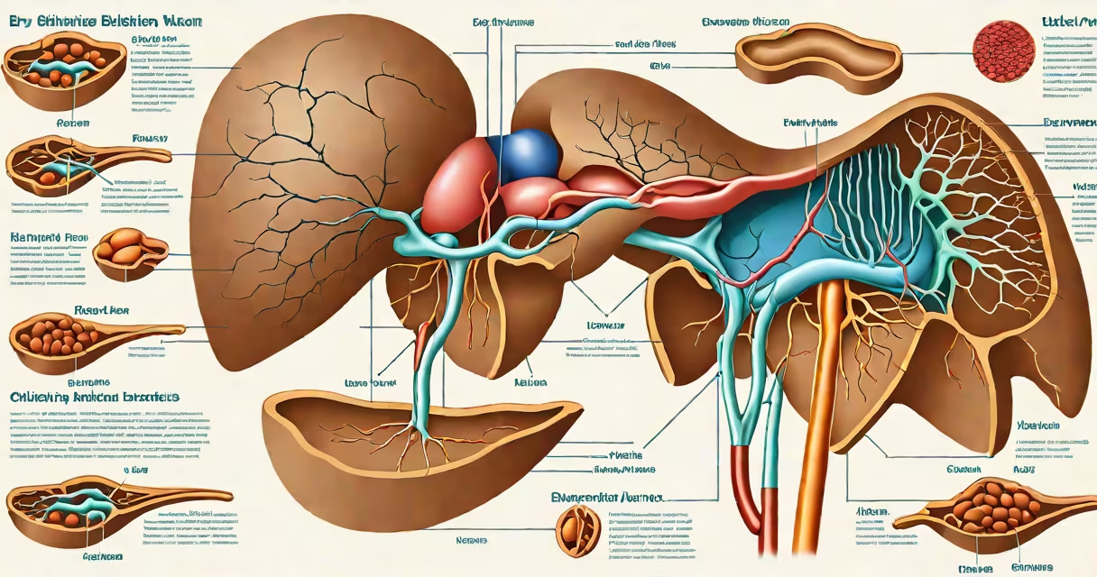
Biliary refers to the system responsible for producing, storing, and transporting bile-the fluid that aids fat digestion and removes waste products from the liver. This system includes the liver, gallbladder, and bile ducts.
Bile is made in the liver's hepatocytes and flows through tiny channels called bile canaliculi into larger ducts. These ducts merge into the common hepatic duct, which joins the cystic duct from the gallbladder to form the common bile duct. The common bile duct empties bile into the duodenum during digestion. When not needed, bile is stored in the gallbladder, where it concentrates up to tenfold.
The term originates from the Latin *bilis*, meaning bile, and *biliaris*, meaning relating to bile. Medical professionals use "biliary" to describe conditions affecting bile flow, such as cholecystitis (gallbladder inflammation) or biliary obstruction. Understanding this term is essential for interpreting diagnoses involving the liver, gallbladder, or bile ducts.
The biliary system functions in three phases: production, storage, and release. The liver synthesizes about 500-1000 mL of bile daily. Bile contains water, electrolytes, cholesterol, phospholipids, and bilirubin-a breakdown product of hemoglobin. Bilirubin gives bile its yellow-green color and is excreted in feces, contributing to stool color.
When food enters the small intestine, especially fatty meals, the hormone cholecystokinin (CCK) signals the gallbladder to contract. This forces concentrated bile into the duodenum via the common bile duct. Bile emulsifies fats, breaking them into smaller droplets so enzymes like lipase can act efficiently. Without bile, fat absorption drops by up to 60%, leading to steatorrhea-fatty, pale stools.
A failure in any part of this process causes clinical symptoms. For example, a gallstone blocking the common bile duct leads to obstructive jaundice-yellowing of skin and eyes due to bilirubin buildup. The condition requires imaging, such as ultrasound or MRCP, to confirm the diagnosis.
The biliary system consists of four primary parts:
| Component | Function | Common Condition |
|---|---|---|
| Liver | Produces bile; metabolizes toxins and drugs | Hepatitis, cirrhosis |
| Gallbladder | Stores and concentrates bile | Cholecystitis, gallstones |
| Common hepatic duct | Carries bile from liver to junction point | Strictures, tumors |
| Common bile duct | Transports bile to duodenum; joins pancreas' duct | Choledocholithiasis, pancreatitis |
Each component plays a distinct role. The liver processes bilirubin and exports it into bile. The gallbladder concentrates bile by absorbing water and electrolytes-this increases bile salt concentration by up to 10 times. The ducts serve as conduits, with the common bile duct sharing a terminal opening with the pancreatic duct at the ampulla of Vater.
When the ampulla becomes blocked-by a stone or tumor-both bile and pancreatic juice cannot exit. This leads to acute pancreatitis, a life-threatening condition requiring immediate intervention. According to the American Gastroenterological Association, biliary pancreatitis accounts for 40% of all acute pancreatitis cases.
"Biliary" is often confused with "hepatic," though they are not interchangeable. Hepatic refers specifically to the liver, while biliary encompasses the entire bile transport network. A patient with hepatitis has liver inflammation; someone with biliary colic experiences pain from bile duct obstruction, even if the liver itself is healthy.
Another misconception is that bile is produced only during meals. In reality, bile flows continuously from the liver. During fasting, bile drains into the duodenum, but most is diverted to the gallbladder for storage. This continuous secretion ensures bile is ready when fat arrives.
Some assume that removing the gallbladder eliminates bile production. That's incorrect. The liver continues making bile, but without storage and concentration, bile flows steadily into the intestines. Most people adapt well, though some develop diarrhea after high-fat meals due to unregulated bile delivery.
A 58-year-old woman presents with jaundice, dark urine, and pale stools. Blood tests show elevated direct bilirubin (4.2 mg/dL) and alkaline phosphatase (180 U/L). An abdominal ultrasound reveals a 1.2 cm stone in the common bile duct.
This case illustrates biliary obstruction. The stone blocks bile flow, causing bilirubin to back up into the bloodstream. The patient's skin and eyes turn yellow-jaundice. Her urine turns dark because conjugated bilirubin is excreted in urine. Stools lose their brown color due to lack of bilirubin in the gut.
Endoscopic retrograde cholangiopancreatography (ERCP) was performed. A sphincterotomy cut the muscle around the ampulla, allowing the stone to be removed. After treatment, her bilirubin dropped to 0.8 mg/dL within 48 hours. This outcome underscores how important the biliary system is-and how quickly dysfunction manifests clinically.
I once assisted in a laparoscopic cholecystectomy. The surgeon identified a gallbladder filled with hundreds of small stones. The organ was inflamed and adherent to surrounding tissue. After removing the gallbladder, we confirmed the cystic duct was patent and free of stones. The patient recovered fully and reported no digestive issues six months later.
This experience reinforced that biliary disease often stems from impaired bile concentration. Stones form when bile becomes supersaturated with cholesterol or calcium bilirubinate. Risk factors include obesity, rapid weight loss, and estrogen therapy. The procedure is routine but highlights how critical the biliary system is-even when it fails.
Understanding what biliary means enables accurate interpretation of medical reports, imaging results, and symptoms. It's not just a medical term-it's a functional system central to digestion and detoxification. Knowing this helps patients and providers alike recognize early signs of disease.