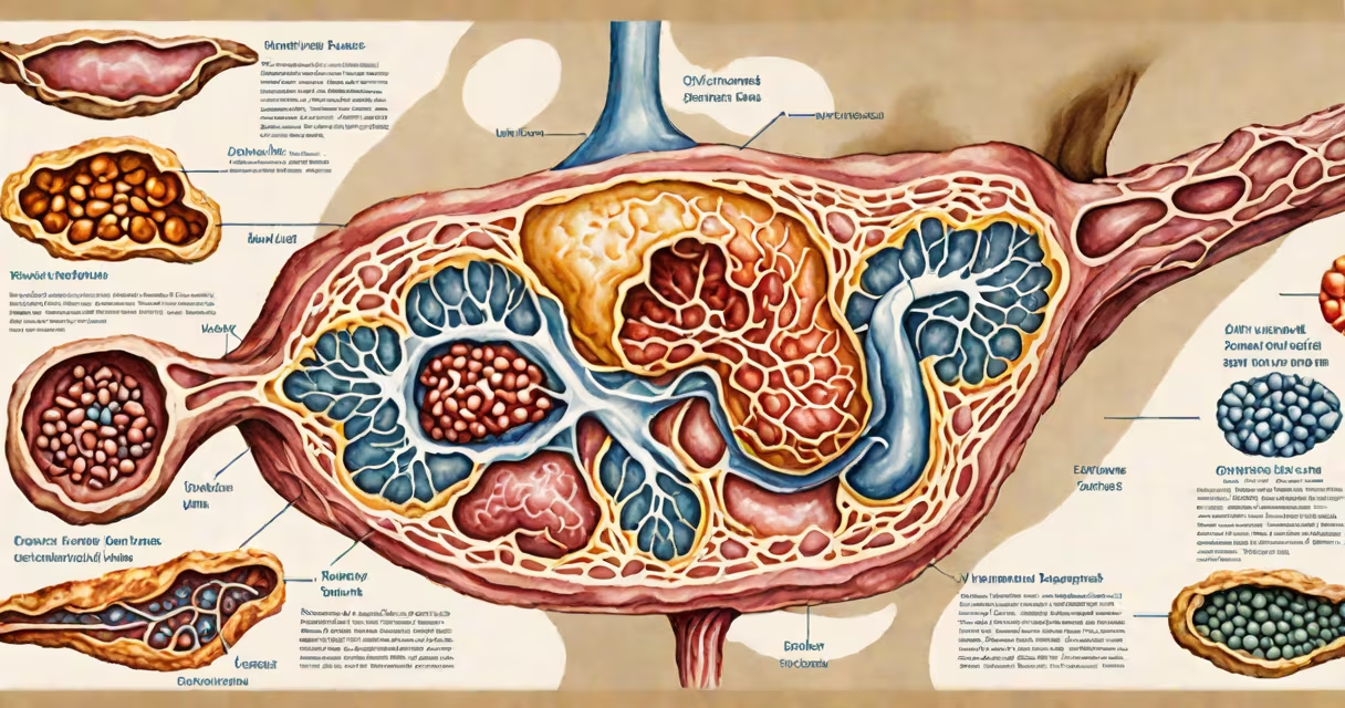
A mass on the pancreas is an abnormal tissue growth identified through imaging such as CT, MRI, or ultrasound. These masses can be benign, precancerous, or malignant, and their significance depends on size, shape, location, and cellular characteristics. Not all masses require immediate treatment-some are monitored over time.
Most pancreatic masses are discovered incidentally during scans done for unrelated reasons. The pancreas lies deep in the upper abdomen, behind the stomach, making symptoms subtle until the mass grows large. When symptoms do appear, they may include upper abdominal pain, unexplained weight loss, jaundice, or new-onset diabetes.
According to the American Cancer Society, about 60,000 cases of pancreatic cancer are diagnosed annually in the U.S., and many begin as small masses detected on imaging. Early detection improves outcomes, but only about 10% of patients survive five years when diagnosed at advanced stages. This underscores the importance of accurate characterization of any pancreatic mass.
Imaging is the primary method for detecting a mass on the pancreas. A contrast-enhanced CT scan or MRI with dedicated pancreatic sequences is standard. These scans show whether the mass is solid, fluid-filled, or mixed. Ultrasound may be used initially, especially if jaundice is present, to assess bile duct obstruction.
Once a mass is seen, radiologists classify it using criteria like size, margins, internal structure, and enhancement patterns. For example, a well-defined, smooth-edged mass less than 2 cm with uniform density is more likely benign. A poorly defined, irregular mass with necrosis or vascular invasion raises concern for malignancy.
The Fukuoka guidelines recommend using the EUS (endoscopic ultrasound)-guided fine needle aspiration (FNA) biopsy when a mass is suspicious for cancer. This procedure allows direct sampling of tissue under real-time imaging, improving diagnostic accuracy. The National Complete Cancer Network (NCCN) notes that biopsy results help determine whether surgery is appropriate or if systemic therapy should be considered.
Not all masses on the pancreas are tumors. They fall into several categories based on composition and behavior:
| Type | Characteristics | Prevalence | Common Management |
|---|---|---|---|
| Solid pseudopapillary neoplasm | Low-grade malignancy, often in young women, well-circumscribed, slow-growing | ~1% of pancreatic tumors | Surgical resection recommended due to risk of bleeding or rupture |
| Neuroendocrine tumor (NET) | Can secrete hormones; varies in grade; low-grade tumors grow slowly | ~10-15% of pancreatic tumors | Resection if localized; targeted therapies if metastatic |
| Pancreatic ductal adenocarcinoma (PDAC) | Most common pancreatic cancer; arises from duct cells; aggressive | ~90% of malignant pancreatic masses | Chemotherapy and radiation; surgery only if early stage |
| Cystic lesions (serous, mucinous, intraductal papillary mucinous neoplasm) | Fluid-filled; some have cancer risk; IPMN can progress to invasive cancer | ~10-20% of detected masses | Monitoring or surgery depending on size and features |
One non-obvious distinction: not all cysts are harmless. Serous cystadenomas are typically benign and rarely progress to cancer. Mucinous cystic neoplasms, however, carry a 10-20% risk of becoming invasive. The presence of mural nodules or thickened walls increases suspicion for malignancy.
I once reviewed a case where a 58-year-old patient had a 3.2 cm cystic lesion in the body of the pancreas. Initial MRI showed no enhancement and no solid components. After follow-up imaging six months later, the lesion grew to 4.1 cm and developed a small nodule. An EUS-FNA confirmed mucinous epithelium with dysplasia-indicating high-grade potential. The patient underwent distal pancreatectomy. Pathology revealed invasive adenocarcinoma. This case illustrates why monitoring and timely intervention matter.
Many people assume any mass on the pancreas is cancer. That's incorrect. Up to 60% of pancreatic masses are benign. The most common benign lesions include serous cystadenomas and insulinomas (tumors that secrete insulin).
Another misconception is that symptoms always precede diagnosis. In reality, 70% of pancreatic masses are asymptomatic at detection. Pain, jaundice, or weight loss usually appear only when the mass exceeds 3 cm or involves nearby nerves or organs.
Doctors sometimes confuse a mass with chronic pancreatitis. Both can cause a focal swelling on imaging. However, pancreatitis typically shows diffuse enlargement, calcifications, and dilated ducts-features absent in isolated masses. Differentiating them prevents unnecessary surgery.
If a mass is found on imaging, the next step is not immediate surgery. Instead, a multidisciplinary team evaluates the lesion. This includes a gastroenterologist, radiologist, surgeon, and oncologist. The decision hinges on size, location, imaging features, and patient health.
For masses under 2 cm with benign features, observation every 6-12 months is standard. Larger or suspicious masses undergo further testing. The Mayo Clinic recommends baseline CT or MRI followed by EUS-FNA if the lesion is ≥2 cm or has worrisome features like irregular borders or lymph node involvement.
Patients with a family history of pancreatic cancer or genetic syndromes like Lynch syndrome or familial adenomatous polyposis should be screened earlier. The National Institute of Diabetes and Digestive and Kidney Diseases (NIDDK) advises annual MRI or EUS for those at high risk.
After diagnosis, treatment plans vary. Surgery is curative only in early-stage disease. For unresectable or metastatic cases, systemic therapy with gemcitabine plus nab-paclitaxel is standard. Clinical trials are available for patients with rare subtypes.
Understanding what is a mass on the pancreas begins with recognizing it as a broad category-not a single condition. Accurate classification determines outcome. A mass is not a death sentence. Many are treatable, even curable, when caught early.
A mass on the pancreas is a clinical finding-not a diagnosis. Its meaning depends on context, imaging, and pathology. Regular screening and prompt evaluation reduce harm and improve survival.