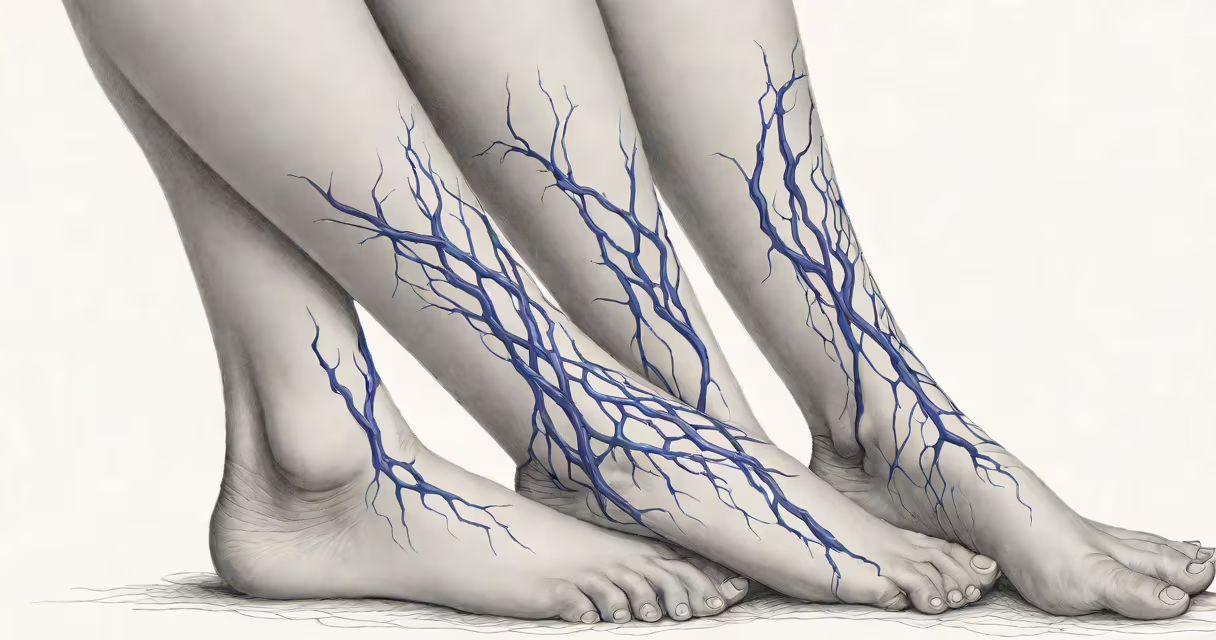
Varicose veins are enlarged, twisted veins that appear near the surface of the skin, most commonly in the legs. They develop when valves inside veins fail, allowing blood to pool and stretch the vessel walls. This condition affects approximately 20-25% of adults in the United States, with women more frequently affected than men.
These veins form due to weakened vein walls and damaged valves. Normally, one-way valves prevent blood from flowing backward. When valves deteriorate, blood accumulates, increasing pressure and causing veins to bulge. The process typically begins in the superficial venous system of the lower limbs, where gravity exacerbates the issue during standing or walking.
Varicose veins are not just cosmetic concerns. They can cause aching, heaviness, itching, and nighttime cramping. In advanced cases, they may lead to skin discoloration, ulcers, or blood clots. According to the American College of Phlebology, about 10% of people with varicose veins develop complications requiring medical intervention.
Veins carry deoxygenated blood back to the heart. In the legs, this requires effort against gravity. Healthy veins rely on muscle contractions and functional valves to maintain forward flow. When valves weaken or fail, blood regurgitates and collects in the veins.
This backup increases intravenous pressure, stretching the vein wall. Over time, the vein loses elasticity and becomes tortuous. The result is a visible, rope-like structure beneath the skin. The process is progressive and worsens with prolonged standing, obesity, or hormonal changes such as those during pregnancy.
A 2021 study published in JAMA Dermatology found that individuals with a family history of varicose veins had a 3.2 times higher risk of developing them compared to those without a family history. This indicates a strong genetic component, even though lifestyle factors also play a role.
Varicose veins are classified by size, appearance, and clinical impact. The most common types include:
| Type | Description | Common Location | Typical Symptoms |
|---|---|---|---|
| Truncal varices | Larger, 3-6 mm diameter veins running along the saphenous system | Inner thigh, behind knee | Pain, swelling, visible bulging |
| Reticular veins | Smaller, bluish networks beneath the skin | Lower leg, above ankle | Often asymptomatic; may itch or burn |
| Saphenous vein reflux | Failure of the great saphenous vein valve, leading to reverse flow | Longitudinal path on inner leg | Heaviness, fatigue after standing |
Severity is assessed using the Clinical, Etiological, Anatomical, and Pathophysiological (CEAP) classification system. Stage C2 involves visible, palpable varicose veins without skin changes. By stage C6, there is active venous ulceration. Most patients present at C2 or C3 (edema).
One non-obvious distinction: varicose veins differ from spider veins (telangiectasias), which are smaller, red or blue threadlike vessels. Spider veins rarely cause symptoms and do not require treatment unless for cosmetic reasons. Varicose veins, however, are associated with measurable hemodynamic dysfunction.
Primary causes include hereditary predisposition, prolonged standing, obesity, and hormonal fluctuations. Pregnancy increases risk due to elevated progesterone, which relaxes vein walls, and increased blood volume. Women who have had multiple pregnancies are twice as likely to develop varicose veins compared to nulliparous women, according to the National Institutes of Health.
Other contributors include aging, lack of physical activity, and wearing high heels regularly. High heels reduce calf muscle pump efficiency-the mechanism that helps push blood upward. A 2019 study in Phlebology showed that women who wore heels over 5 cm daily had a 40% higher incidence of varicose veins over 10 years.
Contrary to popular belief, crossing legs does not cause varicose veins. However, it may temporarily impair circulation. The myth persists because people associate sitting posture with poor venous return, but no evidence links leg-crossing to structural vein damage.
Many confuse varicose veins with other vascular conditions. For example, deep vein thrombosis (DVT) involves clot formation in deep leg veins. DVT presents with sudden swelling, warmth, and pain-often in one leg-and requires urgent treatment. Varicose veins, by contrast, develop slowly and are usually bilateral.
Another frequent mix-up is between varicose veins and lymphedema. Lymphedema results from blocked lymphatic drainage, causing persistent swelling without visible vein distortion. Edema from varicose veins is often intermittent and improves with elevation. A key differentiator: varicose veins show visible, dilated vessels; lymphedema does not.
I once observed a patient who believed her leg swelling was due to varicose veins. After ultrasound imaging, we found she had chronic venous insufficiency with reflux in the saphenous vein. Her symptoms improved after endovenous laser ablation. This case highlights that visible veins don't always correlate with symptom severity-diagnosis requires objective testing.
Diagnosis begins with a physical exam and Doppler ultrasound. This non-invasive test evaluates blood flow direction and valve function. The test detects reflux-backward flow-over 1 second or more, confirming venous insufficiency.
Treatment ranges from conservative to surgical. Conservative measures include compression stockings (20-30 mmHg), regular exercise, and leg elevation. These reduce edema and slow progression. Compression therapy reduces vein diameter by up to 30% in 6 weeks, according to a 2020 randomized trial in Circulation.
For moderate to severe cases, minimally invasive procedures are preferred. Endovenous thermal ablation uses heat via laser or radiofrequency to close diseased veins. Success rates exceed 95% at 5-year follow-up. Sclerotherapy injects a solution into smaller veins, causing scarring and closure. It's effective for reticular and small varicose veins.
Open surgery (stripping) is now rare. It involves removing the entire saphenous vein through incisions. Recovery takes longer, and complication rates are higher. Modern guidelines from the American Venous Forum recommend ablation over stripping for primary treatment.
See a healthcare provider if you experience persistent leg pain, swelling, skin discoloration, or open sores. Early intervention prevents irreversible tissue damage. A 2022 review in Journal of Vascular Surgery found that treating varicose veins before ulcer development reduced healing time by 60%.
Untreated varicose veins may progress to venous stasis dermatitis-a condition marked by brownish pigmentation and thickened skin. If left unchecked, this can evolve into a venous ulcer, which takes months to heal and often recurs.
Varicose veins are a treatable condition rooted in circulatory failure. They are not simply a sign of aging or poor habits. With accurate diagnosis and timely care, symptoms can be managed, progression halted, and quality of life restored.