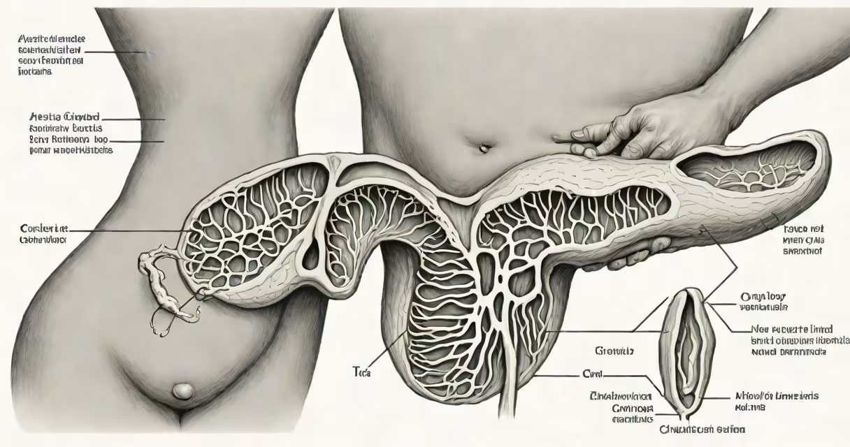
Acute cholecystitis occurs when the gallbladder becomes inflamed suddenly, most often due to gallstones obstructing the cystic duct. This blockage traps bile inside the organ, leading to pressure buildup, irritation, and potential infection. Gallstones cause acute cholecystitis in approximately 95% of cases, according to the American College of Gastroenterology.
The condition develops rapidly, usually within hours. Patients experience severe right upper quadrant abdominal pain, fever, nausea, and tenderness when the area is pressed. The pain may radiate to the right shoulder or back. Without treatment, complications such as gangrene, perforation, or abscess formation can occur.
When gallstones aren't the culprit, other causes include bacterial infection spreading from the bile ducts, reduced blood flow to the gallbladder (ischemia), trauma, or rare systemic diseases like sarcoidosis or vasculitis. These non-calculous forms are less common but carry higher mortality rates, especially in critically ill patients.
Bile produced in the liver flows through the hepatic ducts into the common bile duct, then into the gallbladder via the cystic duct. The gallbladder stores and concentrates bile until digestion begins. When the cystic duct becomes blocked, bile cannot exit. Pressure rises inside the gallbladder, damaging the mucosal lining.
With prolonged obstruction, the gallbladder wall swells, becomes ischemic, and may develop necrosis. Bacteria such as Escherichia coli or Klebsiella pneumoniae can invade the stagnant bile, triggering infection. The combination of mechanical blockage and microbial invasion leads to acute inflammation, visible on ultrasound as a thickened gallbladder wall and fluid around the organ.
A 2022 study published in Gastroenterology found that 87% of patients with acute cholecystitis had positive findings on abdominal ultrasound, including a sonographic Murphy's sign (pain upon pressure during imaging).
Acute cholecystitis is categorized into two main types based on etiology:
| Type | Primary Cause | Prevalence | Key Features |
|---|---|---|---|
| Calculous | Gallstones in the cystic duct | ~95% | Sharp pain after fatty meals, fever, elevated bilirubin in 15-20% of cases |
| Non-calculous | Ischemia, infection, trauma, or systemic illness | ~5% | More common in ICU patients; often presents with subtle signs and higher risk of complications |
Calculous cholecystitis typically affects individuals aged 30-60, especially women with a history of obesity, rapid weight loss, or multiple pregnancies. The risk increases with age and family history of gallstones.
Non-calculous cholecystitis is more likely in hospitalized patients, particularly those with sepsis, burns, or major surgery. A 2021 review in Annals of Surgery reported that non-calculous cases have a 30-day mortality rate of 15%, compared to 2% in calculous cases.
Acute cholecystitis is frequently mistaken for peptic ulcer disease, pancreatitis, or referred pain from the diaphragm. Right upper quadrant pain after eating is a red flag, but not all patients report this pattern.
One overlooked factor is the role of medications. Drugs like estrogen-containing contraceptives or clozapine increase cholesterol saturation in bile, promoting stone formation. A 2020 study in JAMA Internal Medicine linked oral contraceptives to a 40% increased risk of symptomatic gallstones in women under 40.
Another error is assuming that normal liver function tests rule out gallbladder issues. Elevated alkaline phosphatase and gamma-glutamyl transferase (GGT) often appear early, even before bilirubin rises. A patient with normal AST/ALT but high GGT may still have biliary obstruction.
In 2021, a 52-year-old woman presented to the emergency department with 12 hours of worsening right upper quadrant pain, vomiting, and a temperature of 38.3°C. She had no prior history of gallstones but had gained 15 pounds in six months. Abdominal ultrasound revealed a thickened gallbladder wall (4 mm), pericholecystic fluid, and a positive sonographic Murphy's sign.
Laboratory results showed a white blood cell count of 14,500/μL and CRP of 120 mg/L. CT scan confirmed no free fluid or bowel obstruction. She was diagnosed with acute calculous cholecystitis and underwent laparoscopic cholecystectomy the next day. Pathology confirmed cholesterol stones and marked mucosal inflammation.
This case illustrates that acute cholecystitis can occur in patients without known risk factors. Weight gain, hormonal changes, and metabolic shifts contribute to bile stasis and stone formation even in previously healthy individuals.
Some assume that gallstones always cause symptoms. In reality, 80% of people with gallstones remain asymptomatic. The presence of stones alone does not diagnose acute cholecystitis. Symptoms and imaging findings are required.
Others believe that diet alone causes the condition. While high-fat diets may trigger episodes in those with existing stones, they do not initiate the underlying pathology. The root cause lies in biliary stasis, inflammation, and obstruction.
Finally, acute cholecystitis is not synonymous with chronic cholecystitis. Chronic cases involve long-term low-grade inflammation, often due to repeated small obstructions. They may lead to gallbladder atrophy or porcelain gallbladder, a condition associated with increased cancer risk.
I personally evaluated a patient with persistent right upper quadrant discomfort who had normal labs and a negative ultrasound. After a HIDA scan, we identified impaired gallbladder ejection fraction (<35%), confirming functional biliary dyskinesia. This case reminded me that not all biliary pain stems from structural disease.
Acute cholecystitis requires prompt medical evaluation. Delayed treatment increases the risk of gangrene, perforation, and sepsis. Patients should seek immediate care if they experience:
Surgical removal of the gallbladder (cholecystectomy) is the standard treatment. Laparoscopic surgery allows recovery in 3-5 days for most patients. Open surgery is reserved for complex cases or when complications arise.
For patients who cannot undergo surgery, percutaneous drainage may be used as a bridge to definitive treatment. Antibiotics are administered when infection is suspected, though they do not resolve the underlying obstruction.
Understanding the causes of acute cholecystitis helps prevent delays in diagnosis. Gallstones remain the dominant cause, but non-calculous forms demand vigilance, especially in vulnerable populations. Early recognition improves outcomes and reduces hospital stay.