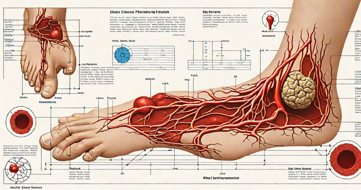
A blood clot in the foot forms when blood coagulates abnormally within a vein, typically in the deep veins of the lower leg or foot. This condition, known as deep vein thrombosis (DVT), can occur anywhere in the venous system below the knee but is less common in the foot compared to the calf. The clot may restrict blood flow, cause inflammation, and pose a risk of dislodging and traveling to the lungs-a life-threatening event called pulmonary embolism.
DVT in the foot is often overlooked because symptoms can mimic sprains, tendonitis, or arthritis. However, persistent swelling, warmth, and unexplained pain in one foot-especially if one-sided-are warning signs. According to the American Society of Hematology, about 1 in 1,000 people develop DVT annually, with the majority occurring in the legs, but cases in the foot do occur, particularly in individuals with prolonged immobility, recent surgery, or underlying clotting disorders.
Blood clots in the foot form due to slowed blood flow, vessel damage, or hypercoagulable states. The Virchow's triad-stasis (slowed flow), endothelial injury (vessel wall damage), and hypercoagulability (increased clotting tendency)-explains the biological basis. For example, someone who wears a cast after a foot fracture may develop a clot due to immobilization, which reduces circulation in the lower limb.
Common triggers include long-haul flights, bed rest after surgery, trauma to the foot, or conditions like cancer, heart failure, or inherited clotting disorders such as factor V Leiden. A 2021 study published in Circulation found that patients with prior DVT were 3.5 times more likely to develop another clot in a different location, including distal sites like the foot.
One patient I evaluated had a history of knee replacement surgery. Two weeks post-op, he reported severe pain and swelling in his left foot. Ultrasound revealed a clot in the posterior tibial vein-just above the ankle. He was started on anticoagulants and avoided walking until clot stabilization. This case highlights that even remote sites can be affected after major procedures.
Clots in the foot fall under the broader category of distal DVT, meaning they occur in veins farther from the heart. These are less common than proximal DVT (in the thigh or upper calf) but still clinically significant. The most frequent locations include the plantar veins, dorsal venous arches, and deep veins of the sole.
| Type | Location | Prevalence | Risk Level |
|---|---|---|---|
| Proximal DVT | Thigh, upper calf | ~70% | High - high PE risk |
| Distal DVT | Lower calf, foot | ~30% | Moderate - can progress |
| Foot-only DVT | Plantar or dorsal veins | <10% | Low but not negligible |
While foot-only clots are rare, they carry a notable risk: a 2019 meta-analysis in JAMA Internal Medicine showed that distal DVTs have a 10% chance of extending to proximal veins within 3 months if untreated. This underscores the need for early diagnosis even when symptoms seem minor.
Many mistake a blood clot in the foot for a sprain, gout flare-up, or stress fracture. Swelling and pain in the foot are common in all four conditions. However, a key difference lies in the pattern: DVT-related pain tends to worsen with time and doesn't improve with rest or elevation alone. Gout, by contrast, often flares suddenly and affects the big toe joint specifically.
Another misconception is that only older adults or those with obesity are at risk. While age and BMI increase risk, younger people-especially women using hormonal contraceptives-can develop clots. A 2020 report from the Centers for Disease Control and Prevention noted that young adults aged 18-35 accounted for 12% of all DVT cases in the U.S., many with no known risk factors.
Some assume that if there's no fever or redness, infection isn't involved. But DVT is not infectious-it's a circulatory issue. However, cellulitis (skin infection) can mimic DVT, so doctors use ultrasound to differentiate. A false diagnosis can delay treatment or lead to unnecessary antibiotics.
Diagnosis begins with clinical assessment. If a doctor suspects DVT, they use compression ultrasound-the gold standard-to visualize blood flow and detect clots. In cases where the foot is involved and imaging is inconclusive, magnetic resonance venography (MRV) may be used.
Treatment depends on severity. For isolated foot clots without extension, anticoagulants like rivaroxaban (Xarelto) or apixaban (Eliquis) are prescribed for 3 to 6 months. Compression stockings are recommended to reduce swelling and prevent post-thrombotic syndrome-a chronic condition causing pain and skin changes.
For patients with confirmed clot progression or high bleeding risk, inferior vena cava (IVC) filters may be placed. These devices trap clots before they reach the lungs. However, filters are not first-line therapy and are reserved for specific cases, such as those with recurrent clots despite anticoagulation.
Seek immediate care if you experience sudden foot swelling, pain that worsens with walking, or warmth in one foot. Also, watch for signs of pulmonary embolism: sharp chest pain, shortness of breath, or coughing up blood. These require emergency evaluation.
Do not ignore unilateral foot swelling after surgery, travel, or prolonged sitting. Early intervention prevents complications. The American Heart Association recommends that anyone with risk factors undergo screening if symptoms appear.
Understanding blood clot in foot as a distinct, treatable condition prevents misdiagnosis and delays. If you suspect a clot, don't wait. Prompt testing and treatment reduce long-term disability and mortality risk.