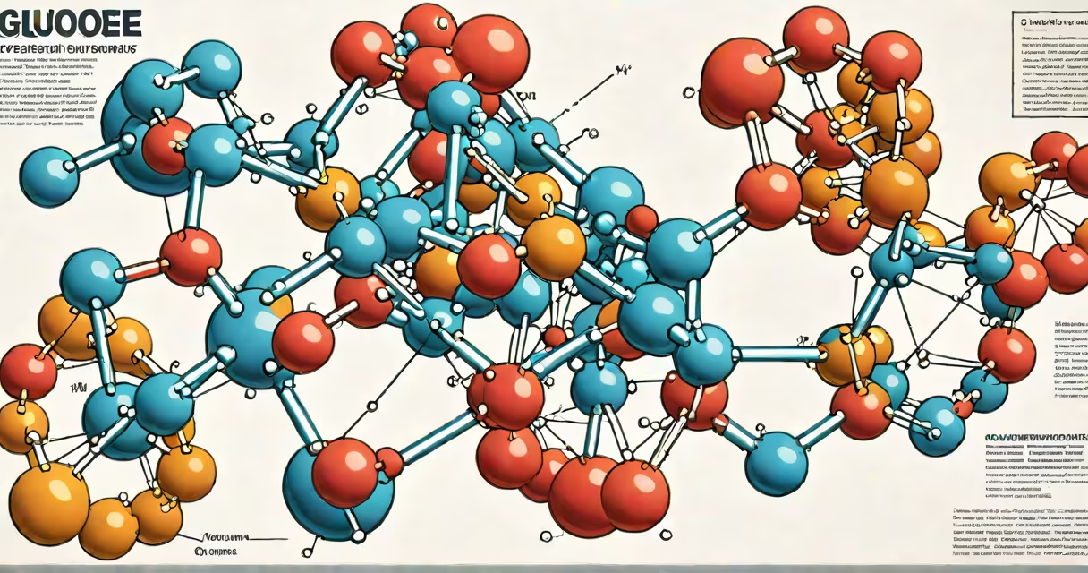
The glucose molecule structure is a six-membered ring composed of five carbon atoms and one oxygen atom, with four hydroxyl groups and one aldehyde group that forms during cyclization. This cyclic form, known as pyranose, is the dominant state in biological systems and determines how enzymes interact with glucose.
Glucose exists primarily as a cyclic hemiacetal due to the reaction between the carbonyl group at carbon 1 and the hydroxyl group on carbon 5. This creates a stable six-membered ring called a pyranose ring. The configuration of the hydroxyl group on carbon 1-called the anomeric carbon-determines whether the molecule is in the alpha or beta form. In the alpha form, the hydroxyl points down (axial) in the standard Haworth projection; in the beta form, it points up (equatorial).
Each carbon in the ring contributes to the molecule's polarity and solubility. The hydroxyl groups make glucose highly water-soluble, enabling transport through blood and cellular fluids. The molecular formula C₆H₁₂O₆ reflects the balance of carbon, hydrogen, and oxygen atoms that allow glucose to serve as a primary energy source in nearly all living organisms.
The precise spatial arrangement of functional groups in the glucose molecule allows it to bind specifically to proteins such as insulin receptors and GLUT transporters. For example, the beta-anomer of glucose fits into the binding site of the GLUT4 transporter in muscle and fat cells, facilitating glucose uptake during insulin signaling.
Enzymes like hexokinase recognize the orientation of hydroxyl groups at carbons 3 and 4. A single misplaced hydroxyl group-such as in galactose-alters how the enzyme binds, preventing phosphorylation and blocking metabolic entry. This specificity helps only correct substrates proceed through glycolysis.
A key distinction often missed: glucose does not exist as a free aldehyde in aqueous solution. The open-chain form accounts for less than 0.02% of circulating glucose. The vast majority is in the cyclic pyranose form, which stabilizes the molecule and prevents unwanted side reactions.
Glucose appears in multiple structural forms depending on conditions. The two most biologically relevant are the alpha-D-glucopyranose and beta-D-glucopyranose isomers. These differ only in the stereochemistry at the anomeric carbon.
| Form | Anomeric OH Orientation | Stability | Biological Role |
|---|---|---|---|
| Alpha-D-glucopyranose | Down (axial) | Less stable | Forms glycogen and starch linkages |
| Beta-D-glucopyranose | Up (equatorial) | More stable | Found in cellulose and bacterial cell walls |
When glucose polymerizes, the linkage type depends on the anomeric configuration. Alpha-1,4-glycosidic bonds connect glucose units in starch and glycogen, allowing rapid breakdown during energy demand. Beta-1,4-glycosidic bonds form cellulose, which humans cannot digest due to lack of beta-glucosidase enzymes.
Non-reducing sugars like sucrose contain glucose linked via its anomeric carbon to another sugar (fructose), locking the ring and preventing further ring-opening. This prevents the molecule from acting as a reducing agent in tests like Benedict's assay.
Many assume glucose is a linear chain. In reality, the open-chain form is transient and unstable in water. The cyclic form dominates even in blood plasma, where glucose concentrations average 5 mmol/L. The equilibrium favors the cyclic structure by a factor of 10⁴.
Another error: thinking all glucose is the same regardless of stereochemistry. D-glucose and L-glucose are enantiomers. Only D-glucose is metabolized by human enzymes. L-glucose is not recognized by glycolytic pathways and passes through the body unchanged.
Even within D-glucose, the difference between alpha and beta anomers matters. During digestion, enzymes like amylase cleave alpha-1,4 bonds but ignore beta linkages. This explains why we can break down starch but not cellulose-even though both are glucose polymers.
Consider a fasting blood glucose test. A result of 110 mg/dL indicates hyperglycemia. This value measures total glucose concentration, including both anomers. However, the clinical relevance lies in the molecule's structure: only the cyclic form is transported into cells via GLUT transporters.
When a lab uses a glucose oxidase assay, it detects the aldehyde form generated during ring opening. The enzyme selectively binds the open-chain intermediate, converting glucose to glucono-δ-lactone. This reaction is proportional to glucose concentration and forms the basis of home glucose meters.
I once observed a patient with unexplained fatigue whose blood glucose was normal, yet symptoms persisted. A follow-up test revealed low intracellular glucose despite normal serum levels. Further analysis showed impaired GLUT1 function due to a mutation affecting the protein's ability to recognize the hydroxyl group orientation on carbon 2 of glucose.
The glucose molecule structure defines its biochemical behavior. Its ring form, stereochemistry, and anomeric state determine how it is absorbed, stored, and broken down. Without this precise structure, life as we know it could not sustain energy metabolism.
Understanding the glucose molecule structure reveals why small changes in atomic arrangement lead to vastly different biological outcomes-from energy storage to disease pathology. This knowledge underpins diagnostics, drug design, and metabolic research.