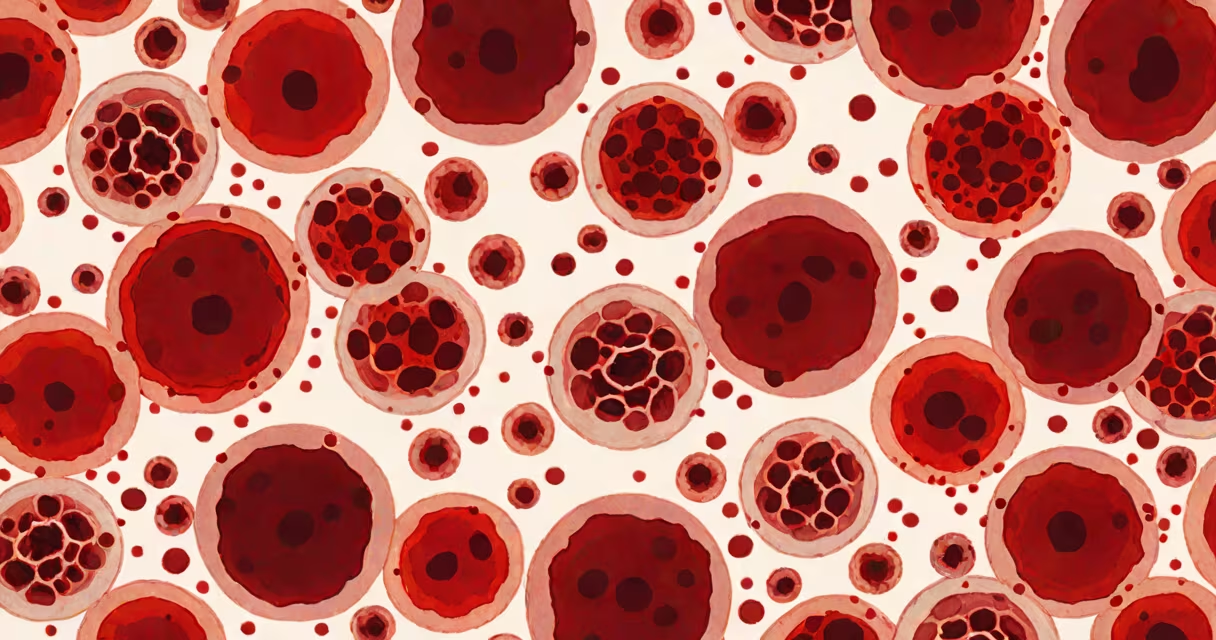
A high red blood cell (RBC) count, also known as erythrocytosis, occurs when the body produces too many red blood cells, increasing blood viscosity and raising the risk of clotting. This condition may result from physiological adaptation, underlying disease, or genetic disorders.
RBCs carry oxygen from the lungs to tissues. A normal adult range is 4.7 to 6.1 million cells per microliter for men and 4.2 to 5.4 million for women. Values above these thresholds indicate elevated RBCs. The most common cause is relative polycythemia due to dehydration, which increases concentration without actual excess production. True polycythemia involves increased RBC mass and requires investigation.
The bone marrow produces red blood cells under the influence of erythropoietin (EPO), a hormone released by the kidneys in response to low oxygen levels. When tissue oxygenation drops-due to lung disease, high altitude, or heart failure-the kidneys secrete more EPO, stimulating RBC production. This compensatory mechanism maintains oxygen delivery but can lead to excessive RBC counts if sustained.
In primary polycythemia vera, a mutation in the JAK2 gene leads to uncontrolled RBC production independent of EPO. This rare disorder affects about 2 in 10,000 people and increases the risk of thrombosis, hemorrhage, and progression to myelofibrosis or acute leukemia. Diagnosis requires testing for the JAK2 V617F mutation and exclusion of secondary causes.
Secondary polycythemia results from external stimuli. Chronic obstructive pulmonary disease (COPD) reduces oxygen exchange, triggering persistent EPO release. Smoking, which damages alveoli and promotes hypoxia, contributes to this pattern. Living at high altitudes also elevates RBCs as a natural adaptation to lower atmospheric oxygen.
Conditions leading to high RBCs fall into two categories: secondary (reactive) and primary (clonal). Secondary causes are more frequent and often reversible. Primary polycythemia vera is less common but carries higher long-term risks.
| Type | Key Causes | Diagnostic Clue |
|---|---|---|
| Secondary Polycythemia | Chronic hypoxia (COPD, sleep apnea), high-altitude residence, smoking, renal tumors producing EPO | Elevated serum EPO levels |
| Primary Polycythemia Vera | JAK2 V617F mutation, clonal hematopoiesis | Low or normal EPO despite high RBCs |
| Relative Polycythemia | Dehydration, fluid loss, burns | Normal RBC mass, elevated concentration due to reduced plasma volume |
Dehydration accounts for up to 30% of apparent high RBC counts in clinical settings. A patient presenting with a measured RBC of 6.8 million/µL may have normal RBC mass but reduced plasma volume. Rehydration typically resolves the lab abnormality without treatment.
Many assume a high RBC count always indicates a serious blood disorder. In reality, transient elevations are common and benign. For example, athletes training at high altitude often show elevated RBCs due to acclimatization-a normal adaptive response. These values do not require intervention unless symptoms develop.
Another misconception is equating high RBCs with iron overload. While both conditions can coexist, they are distinct. Iron deficiency leads to low RBCs and hemoglobin. High RBCs with normal iron stores suggest a different etiology. A 2021 study in Haematologica found that 15% of patients initially diagnosed with polycythemia vera had secondary causes upon re-evaluation, underscoring the need for thorough workup.
One non-obvious distinction: high RBCs from smoking or COPD are often accompanied by elevated hemoglobin and hematocrit. However, in polycythemia vera, platelet counts and white blood cell counts may also rise, reflecting pan-myeloproliferation. This triad-increased RBCs, WBCs, and platelets-is a red flag for clonal disease.
A 58-year-old man presented with headaches and dizziness. His CBC showed RBC 6.9 million/µL, hemoglobin 19.2 g/dL, and hematocrit 57%. Initial suspicion was polycythemia vera. But serum EPO was 18 mU/mL-above the normal range of 4-20 mU/mL-indicating a reactive process. Further history revealed he smoked two packs daily and had untreated sleep apnea. Overnight oximetry confirmed nocturnal desaturation. After starting CPAP therapy and quitting smoking, his RBC count dropped to 5.3 million/µL within six weeks.
This case illustrates that high RBCs aren't always pathological. Addressing the root cause-hypoxia from sleep apnea and tobacco exposure-resolved the abnormality without medication.
If a high RBC count persists after hydration or resolving reversible factors, further evaluation is needed. The American Society of Hematology recommends checking serum EPO, JAK2 mutation status, and arterial blood gas analysis. A venous blood gas can reveal chronic respiratory acidosis, indicating long-standing lung disease.
Imaging such as CT angiography may be required if a renal tumor is suspected. Renal cell carcinoma can secrete EPO, mimicking secondary polycythemia. A 2020 review in Blood Advances reported that 1 in 12 cases of unexplained polycythemia were linked to renal masses.
For individuals with a family history of polycythemia vera, genetic counseling and testing for JAK2 mutations are advised. Early detection allows monitoring for complications like thrombosis or transformation to acute leukemia.
As a clinician, I've seen patients misdiagnosed with polycythemia vera based on isolated lab findings. One patient was prescribed phlebotomy and hydroxyurea until we discovered his high RBCs stemmed from severe dehydration during a heatwave. After rehydration, his values normalized. This case reinforced the principle: always assess context before labeling a lab abnormality.
Understanding what would cause RBC to be high begins with distinguishing between true polycythemia, secondary responses, and laboratory artifacts. Dehydration, chronic hypoxia, and genetic mutations are the core drivers. Proper diagnosis prevents unnecessary treatment and identifies treatable conditions early.