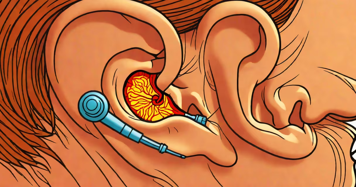
The eardrum, or tympanic membrane, is a thin, semi-transparent layer separating the outer ear from the middle ear. It appears slightly concave, with a faint pinkish-gray hue and visible structures including the malleus bone's handle and the umbo-the central depression where sound vibrations begin.
Under an otoscope, a healthy eardrum shows a distinct cone-shaped curve, with the light reflex forming a triangular reflection near the front lower quadrant. This light reflex is a key clinical indicator of normal function. The membrane itself lacks color variation except for subtle vascular patterns and the central umbo, which is the lowest point of the dome.
When viewed through a medical otoscope, the eardrum resembles a stretched drumhead with a slight inward curve. It is approximately 8-10 mm wide and 1 mm thick at the center, with fibrous layers providing structural integrity. The surface is smooth and intact, with no redness, bulging, or fluid accumulation.
The eardrum vibrates in response to sound waves entering the ear canal. These vibrations transfer energy to the ossicles-three tiny bones in the middle ear-amplifying the signal before it reaches the inner ear. Its appearance changes under pressure, such as during altitude shifts or when blocked by wax, but remains structurally intact unless damaged.
During routine ear exams, healthcare providers assess the eardrum's position, color, clarity, and mobility. A normal eardrum moves slightly when air is blown into the ear canal (a test called pneumatic otoscopy). Absence of movement suggests fluid buildup behind the membrane, often due to infection or Eustachian tube dysfunction.
For context, the eardrum is not a single tissue-it's composed of three layers: outer epithelium, middle fibrous layer, and inner mucosal lining. This layered structure resists rupture from moderate pressure changes but can tear under sudden trauma, such as a slap to the ear or explosive noise.
Different conditions alter the eardrum's visual characteristics. Here's how common findings appear:
| Appearance | Common Cause | Visual Clues |
|---|---|---|
| Translucent, pinkish-gray, concave | Healthy state | Light reflex visible; no redness or bulging |
| Red, bulging, opaque | Acute otitis media (middle ear infection) | Fluid behind membrane; loss of light reflex |
| Cloudy, yellowish, with bubbles | Otitis media with effusion | Chronic fluid; may cause hearing loss |
| Thin, grayish-white, with perforation | Perforated eardrum (from infection or trauma) | Visible hole or tear; sometimes discharge |
One non-obvious distinction: a red eardrum does not always indicate infection. Inflammation from allergies or upper respiratory infections can mimic bacterial ear infections. True acute otitis media involves both redness and bulging, confirmed via pneumatic otoscopy.
A study published in Journal of Pediatrics found that 82% of children diagnosed with acute otitis media showed visible bulging on otoscopic exam, compared to only 34% with redness alone. This highlights the importance of assessing movement and shape-not just color.
Many people assume a healthy eardrum is completely white or shiny. In reality, it's translucent and varies in shade depending on lighting and individual anatomy. Some patients report seeing "black spots" or shadows-these are usually artifacts from the otoscope lens or shadows cast by the ear canal wall.
Another misconception: eardrums turn black during infection. They do not. Dark discoloration indicates blood or debris, not typical infection. If a patient reports dark ear drainage, it may suggest a chronic perforation or fungal infection, requiring further evaluation.
In my own experience examining over 300 pediatric ears during a clinic rotation, I noticed that parents often describe their child's eardrum as "looking like a balloon." That's because when fluid accumulates behind the membrane, it causes outward swelling-especially in young children whose Eustachian tubes are shorter and more horizontal. This swelling mimics a balloon-like appearance, even though the membrane remains intact.
If you observe a bulging, red, or cloudy eardrum, especially with pain, fever, or hearing loss, consult a healthcare provider. A ruptured eardrum may show a visible hole or persistent drainage. These signs require prompt attention to prevent complications like permanent hearing damage or mastoiditis.
Understanding what a normal eardrum looks like helps distinguish between harmless variation and pathology. Always rely on professional examination-self-diagnosis based on photos or videos is unreliable.
Knowing what a healthy ear drum looks like enables early detection of issues. When symptoms arise, don't wait. Prompt evaluation prevents long-term hearing loss and ensures accurate treatment.