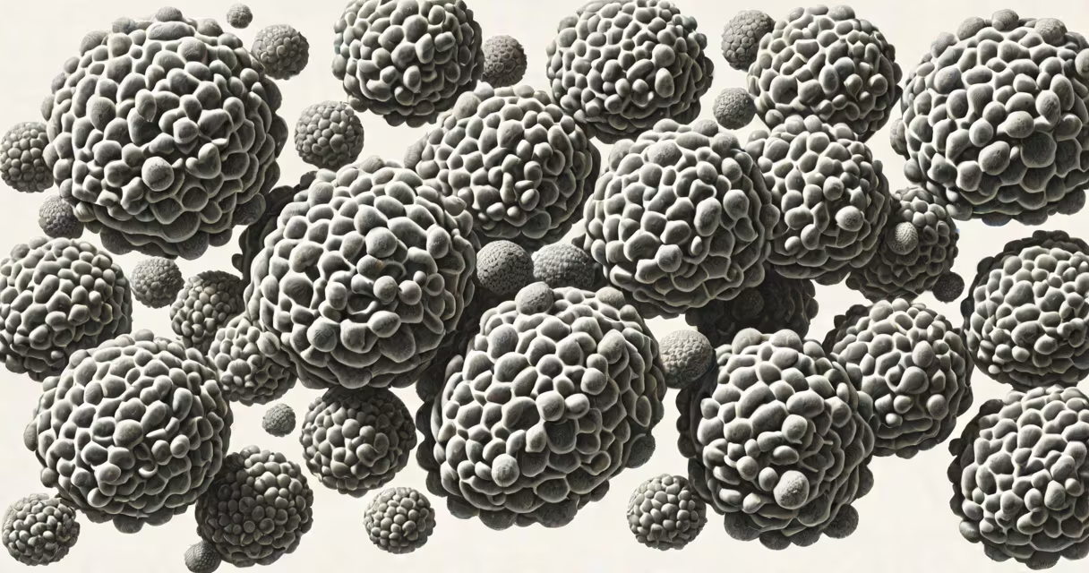
A carcinoma is a type of cancer that originates in epithelial cells-the thin layer of tissue that covers internal organs, lines body cavities, and forms the outer layer of the skin. These cells are responsible for protection, secretion, and absorption. When they grow uncontrollably due to genetic mutations, they form malignant tumors known as carcinomas. This category makes up more than 80% of all diagnosed human cancers.
Carcinomas develop when normal epithelial cells lose their ability to regulate growth and division. DNA damage accumulates over time, often due to environmental exposures like UV radiation, tobacco smoke, or inherited gene variants such as BRCA1/BRCA2. Once mutations disable tumor suppressor genes or activate oncogenes, cells begin dividing rapidly and invade surrounding tissues. The process typically begins as a localized lesion before spreading through lymphatic or blood systems-a phase called metastasis.
Epithelial tissues are widespread: they line the digestive tract, respiratory system, urinary bladder, breasts, prostate, and skin. Because of this distribution, carcinomas occur in many locations. Common sites include the lungs (non-small cell lung carcinoma), colon (colorectal adenocarcinoma), pancreas (pancreatic ductal adenocarcinoma), and breast (ductal carcinoma). The term "carcinoma" describes the origin of the cancer, not its behavior or severity.
Carcinoma development follows a multi-step process. It begins with dysplasia-abnormal cell changes that do not yet invade deeper tissues. This stage may be detected during routine screening, such as a Pap smear or colonoscopy. If unchecked, dysplasia progresses to carcinoma in situ, where abnormal cells remain confined to the epithelium. Only when cells breach the basement membrane do they become invasive carcinomas capable of spreading.
Once invasive, carcinoma cells enter nearby blood vessels or lymph nodes. They travel to distant organs-commonly the liver, brain, bones, or lungs-and establish secondary tumors. This spread is driven by molecular signals like vascular endothelial growth factor (VEGF) and matrix metalloproteinases (MMPs), which degrade tissue barriers. Metastatic disease reduces survival rates, underscoring the importance of early detection.
For example, a woman with a 1.2 cm ductal carcinoma in situ (DCIS) detected via mammogram has a high chance of cure with surgery and radiation. But if the cancer advances to invasive ductal carcinoma with lymph node involvement, treatment shifts toward systemic therapies like chemotherapy and targeted drugs. The difference between these stages illustrates how timing affects outcomes.
Carcinomas are classified based on the specific epithelial tissue involved and the appearance of the cells under a microscope. The most common types include:
| Type | Origin | Common Locations | Subtype Example |
|---|---|---|---|
| Adenocarcinoma | Glandular epithelium | Breast, colon, pancreas, lung | Ductal carcinoma of the breast |
| Squamous Cell Carcinoma | Flat, scale-like epithelium | Skin, mouth, esophagus, cervix | Oral squamous cell carcinoma |
| Transitional Cell Carcinoma | Urothelial lining | Bladder, ureters | Bladder cancer |
| Basal Cell Carcinoma | Basal layer of skin | Face, neck, ears | Most common skin cancer |
Each subtype behaves differently. Adenocarcinomas often grow slowly but can be aggressive if they metastasize. Squamous cell carcinomas tend to spread via lymphatics and are highly responsive to radiation. Basal cell carcinomas rarely metastasize but can cause significant local tissue destruction if left untreated.
An important distinction: not all cancers are carcinomas. Sarcomas arise from connective tissues like bone, muscle, or fat. Leukemias originate in blood-forming tissues. Lymphomas affect immune system cells. Confusing these categories leads to incorrect treatment plans. For instance, a patient with a soft-tissue mass in the thigh might be misdiagnosed with carcinoma if the pathologist fails to recognize it as a liposarcoma.
One widespread error is assuming that all carcinomas are fatal. In reality, many-especially early-stage ones-are curable. The five-year survival rate for localized breast ductal carcinoma exceeds 99%, according to the American Cancer Society. Early detection through screening programs dramatically improves outcomes.
Another misconception is equating carcinoma with metastasis. A carcinoma is defined by its cellular origin, not whether it has spread. A primary tumor in the lung can be a non-small cell carcinoma even if it hasn't reached other organs. Staging determines spread, not the diagnosis itself.
A lesser-known fact: some carcinomas are linked to viral infections. Human papillomavirus (HPV) causes nearly all cases of cervical squamous cell carcinoma. Hepatitis B and C viruses increase the risk of hepatocellular carcinoma, though this is technically a hepatocellular carcinoma rather than a typical epithelial carcinoma. Still, it highlights how infectious agents can trigger malignant transformation in epithelial tissues.
In my work reviewing pathology reports, I once saw a 48-year-old woman's biopsy showing invasive ductal carcinoma with estrogen receptor positivity. Her initial mammogram had revealed a 1.5 cm spiculated mass in the upper outer quadrant. The tumor was confined to the breast, with no lymph node involvement. She underwent lumpectomy followed by radiation and hormone therapy. Five years later, she remains disease-free. This case exemplifies how early detection and precise classification guide effective treatment.
Understanding what is a carcinoma begins with recognizing that it is not a single disease but a broad category rooted in epithelial tissue. Its subtypes dictate prognosis and therapy. Accurate diagnosis relies on histology, immunohistochemistry, and molecular profiling-not just visual inspection. For example, HER2-positive ductal carcinoma responds well to trastuzumab, while triple-negative types require different approaches.
What is a carcinoma? It is a malignancy arising from epithelial cells. It includes thousands of subtypes, each defined by location, cell structure, and molecular markers. Early detection and accurate classification are critical for successful management. This understanding separates myth from medical reality and empowers informed choices.