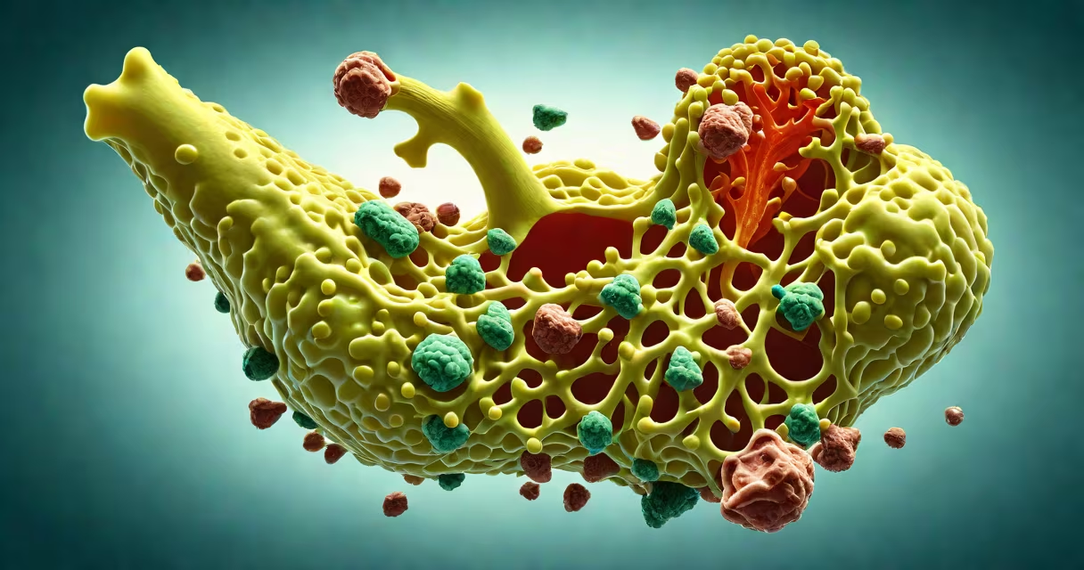
Cholecystitis is inflammation of the gallbladder, typically caused by obstruction of the cystic duct by gallstones. Cholelithiasis refers to the presence of stones in the gallbladder. Together, they represent a common cause of abdominal pain and require medical evaluation.
These conditions frequently occur together. Gallstones form when substances in bile-primarily cholesterol or bilirubin-crystallize and aggregate. When a stone blocks the cystic duct, bile cannot drain properly, leading to pressure buildup and inflammation. This process triggers cholecystitis. Not everyone with gallstones develops symptoms, but when they do, it's often due to cholecystitis.
The American College of Gastroenterology reports that about 10-15% of adults in the U.S. have gallstones, but only 1-4% develop symptomatic cholecystitis annually. The risk increases with age, obesity, female sex, and rapid weight loss. Left untreated, cholecystitis can progress to complications such as gangrene, perforation, or sepsis.
Bile is produced in the liver and stored in the gallbladder. When fat enters the small intestine, the gallbladder contracts and releases bile through the common bile duct into the duodenum. This process aids digestion. Blockage at any point disrupts normal function.
When a gallstone lodges in the cystic duct-the narrow channel connecting the gallbladder to the common bile duct-it prevents bile from exiting. Pressure rises inside the gallbladder. This mechanical obstruction causes ischemia (reduced blood flow), tissue damage, and activation of inflammatory pathways. Neutrophils infiltrate the gallbladder wall, leading to swelling, pain, and fever.
A 2021 study in Gastroenterology found that 85% of patients with acute cholecystitis had gallstones visible on ultrasound. The remaining 15% had sludge or rare causes like tumors or parasites. This confirms that cholelithiasis is the primary driver of acute cholecystitis in clinical practice.
Cholecystitis is classified by duration and severity. Acute cholecystitis develops rapidly-within hours to days-and presents with severe right upper quadrant pain, fever, and positive Murphy's sign (tenderness when the abdomen is pressed under the ribcage during inhalation).
Chronic cholecystitis results from repeated episodes of acute inflammation. Over time, the gallbladder thickens, loses elasticity, and may shrink. It often causes mild, recurrent pain after eating fatty meals. Diagnosis relies on imaging and symptom history.
| Type | Onset | Primary Cause | Common Symptoms |
|---|---|---|---|
| Acute | Sudden (hours) | Gallstone obstruction (95%) | Severe RUQ pain, fever, nausea |
| Chronic | Recurrent over months/years | Repeated acute episodes | Mild bloating, postprandial discomfort |
| Acute acalculous | Sudden, no stones | Severe illness, trauma, surgery | High mortality; requires ICU care |
Cholelithiasis itself has subtypes based on composition. Cholesterol stones make up 80-85% of cases. They form when bile contains too much cholesterol relative to bile salts and lecithin. Pigment stones-accounting for 15-20%-develop in people with hemolytic disorders or cirrhosis, where excess bilirubin leads to stone formation.
One non-obvious fact: not all gallstones cause symptoms. A 2019 study in Annals of Internal Medicine showed that 80% of people with gallstones detected incidentally on imaging remain asymptomatic over 10 years. This challenges the idea that gallstones always require treatment.
Cholecystitis and cholelithiasis are most common in adults aged 40-60. Women are affected twice as often as men-likely due to estrogen's role in increasing cholesterol saturation in bile. Risk factors include pregnancy, oral contraceptives, obesity, and rapid weight loss.
Geographic variation exists. Prevalence is higher in North America and Western Europe, affecting 10-15% of adults. In contrast, rates in parts of Africa and Asia are lower-around 3-5%. This difference correlates with diet, body mass index, and genetic factors influencing bile composition.
I once observed a 58-year-old woman in a clinic who reported intermittent right upper abdominal pain for six months. Ultrasound revealed multiple gallstones but no signs of inflammation. She was asymptomatic between episodes. After discussion, we chose observation over surgery. She remained stable for five years-proof that not all cholelithiasis requires intervention.
Many confuse cholecystitis with pancreatitis. Both involve upper abdominal pain and elevated amylase/lipase enzymes. However, pancreatitis pain radiates to the back and worsens after eating. Cholecystitis pain stays localized to the right upper quadrant and often improves after vomiting.
Another frequent mix-up: biliary colic vs. cholecystitis. Biliary colic is the sharp, cramping pain from a stone temporarily blocking the cystic duct. It lasts 1-6 hours and resolves when the stone moves. Cholecystitis involves persistent inflammation lasting longer than 6 hours and includes fever and lab abnormalities.
Some assume all gallstones need removal. But guidelines from the American Society for Gastrointestinal Endoscopy state that elective cholecystectomy is recommended only for symptomatic patients. Asymptomatic gallstones are managed conservatively unless there's a high-risk condition-such as diabetes or prior gallstone pancreatitis.
Ultrasound is the gold standard for diagnosing both conditions. It detects gallstones with 95% sensitivity and identifies gallbladder wall thickening or pericholecystic fluid-signs of inflammation. CT scans are less sensitive but useful if complications like abscesses or perforation are suspected.
Treatment depends on severity. Acute cholecystitis typically requires hospitalization. Patients receive IV fluids, antibiotics (e.g., ceftriaxone plus metronidazole), and pain control. Surgery-laparoscopic cholecystectomy-is performed within 72 hours of diagnosis. Delaying surgery increases complication rates.
For chronic cholecystitis or recurrent biliary colic, laparoscopic cholecystectomy remains the definitive treatment. Recovery takes 1-2 weeks. Most patients resume normal diets within a month, though some report temporary diarrhea or bloating due to altered fat digestion.
Non-surgical options exist but are limited. Ursodeoxycholic acid can dissolve small cholesterol stones-but only in 30% of patients and takes months to work. It's rarely used today due to low efficacy and compliance issues.
When gallstones are present but no inflammation is detected, management focuses on symptom monitoring. Patients are advised to avoid high-fat meals and maintain a healthy weight. Understanding the underlying causes of acute cholecystitis helps guide prevention and early intervention.
Cholecystitis and cholelithiasis are distinct but interrelated conditions. Cholelithiasis describes the presence of gallstones. Cholecystitis is the resulting inflammation. Accurate diagnosis hinges on clinical presentation, imaging, and lab tests. Not all gallstones lead to symptoms. Not all symptomatic cases require surgery. Proper evaluation avoids unnecessary procedures while preventing serious complications.