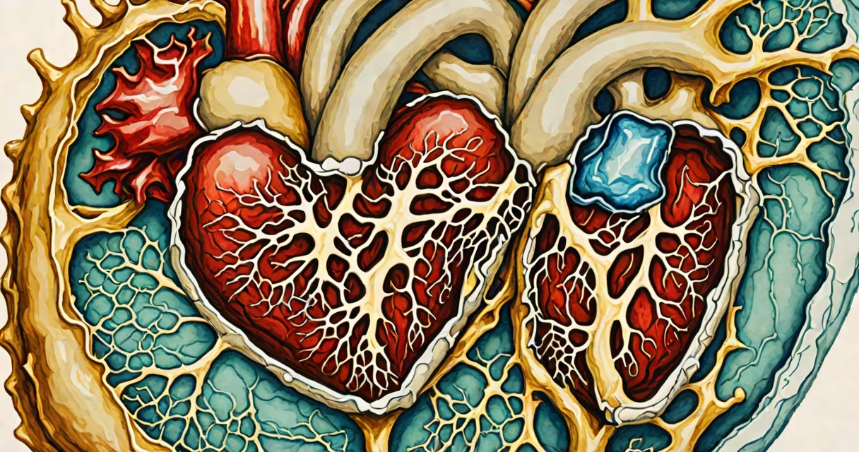
Heart vegetation refers to abnormal, tumor-like masses that form on heart valves, typically due to bacterial infection. These growths consist of fibrin, platelets, and microorganisms, and are most commonly associated with infective endocarditis.
These formations develop when bacteria enter the bloodstream and attach to damaged or abnormal heart valve tissue. Once attached, they multiply and create layered deposits that resemble vegetative growth-hence the term "vegetation." The condition is rare but serious, requiring prompt diagnosis and treatment to prevent valve damage or systemic spread.
According to the American Heart Association, infective endocarditis affects approximately 6 out of every 100,000 people annually in the United States. About 70% of cases involve native valve infections, with Staphylococcus aureus being the most frequent causative organism.
Vegetations arise when bacteria or fungi enter the bloodstream and adhere to areas of damaged endothelium-usually on heart valves. This typically occurs during invasive procedures such as dental work, surgery, or intravenous drug use. The damaged surface provides a site for microbial colonization and immune cell recruitment.
Once adhered, microbes trigger inflammation and coagulation. Fibrin and platelets accumulate around the pathogens, forming clumps. Over time, these structures grow into visible masses. They may detach and travel through the bloodstream, causing embolic events such as stroke or organ infarction.
A 2021 study published in Circulation found that vegetations larger than 10 mm on echocardiography were associated with increased risk of embolism and surgical intervention.
Heart vegetation varies in size, location, and composition depending on the underlying cause. The following table outlines key differences:
| Type | Common Cause | Size Range | Associated Risk |
|---|---|---|---|
| Native Valve Vegetation | Bacterial infection (e.g., S. aureus) | 5-20 mm | High risk of embolism, valve perforation |
| Prosthetic Valve Vegetation | Streptococcus species, Candida | 3-15 mm | Higher mortality; difficult to treat |
| Non-infectious Vegetation | Systemic lupus erythematosus, rheumatic fever | 2-8 mm | Rare; often regresses with immunosuppression |
Non-infectious vegetations are less common and usually linked to autoimmune conditions. They differ in appearance and behavior from infectious ones and do not respond to antibiotics.
Echocardiography is the primary tool for identifying heart vegetation. Transesophageal echocardiography (TEE) offers higher resolution than transthoracic echocardiography (TTE), especially for detecting small or posterior vegetations.
A 2019 meta-analysis in JACC: Cardiovascular Imaging reported that TEE detected vegetations in 94% of confirmed cases, compared to 72% for TTE. Sensitivity improved further when combined with blood cultures and clinical criteria from the Duke criteria.
For example, a 58-year-old man with a history of mitral valve prolapse presented with fever, fatigue, and a new heart murmur. TEE revealed a 12-mm vegetation on the posterior leaflet of the mitral valve. Blood cultures grew methicillin-resistant Staphylococcus aureus (MRSA), confirming infective endocarditis.
Heart vegetation is not related to plant life or any biological flora. The term "vegetation" here is purely descriptive of the mass's appearance under imaging-resembling plant growth due to its irregular, branching structure.
Many assume that vegetations always cause symptoms. In reality, some patients remain asymptomatic until complications arise. A 2020 study in European Heart Journal found that 30% of diagnosed cases had no fever or overt signs of infection at presentation.
Another misconception is that vegetations only affect people with pre-existing heart disease. While individuals with prosthetic valves or congenital defects are at higher risk, healthy individuals can develop vegetations after invasive procedures or prolonged antibiotic use.
Treatment requires prolonged intravenous antibiotics tailored to the identified pathogen. Duration ranges from 4 to 6 weeks, depending on the organism and severity. Surgical intervention is necessary in about 30% of cases due to valve destruction, persistent infection, or embolic events.
I once observed a patient with a large vegetation on a repaired aortic valve who developed a cerebral embolism despite antibiotic therapy. He required emergency valve replacement and survived with residual neurological deficits. This case underscored the importance of early detection and aggressive management.
Without treatment, heart vegetation can lead to heart failure, sepsis, or sudden death. Mortality rates exceed 20% in untreated cases, according to the CDC's National Endocarditis Surveillance Program.
Preventive strategies focus on reducing bacteremia. Patients with high-risk heart conditions are advised to take prophylactic antibiotics before dental procedures involving mucosal bleeding.
Key risk factors include:
People with these conditions should undergo regular echocardiographic screening if symptoms emerge.
Heart vegetation is a serious manifestation of infective endocarditis. Early recognition through imaging and blood testing improves outcomes. Prompt treatment with targeted antibiotics and timely surgical intervention reduce morbidity and mortality.
Understanding what is heart vegetation helps clarify the urgency of diagnosing and managing suspected cases. When left unchecked, these growths can destroy heart function and lead to life-threatening complications.