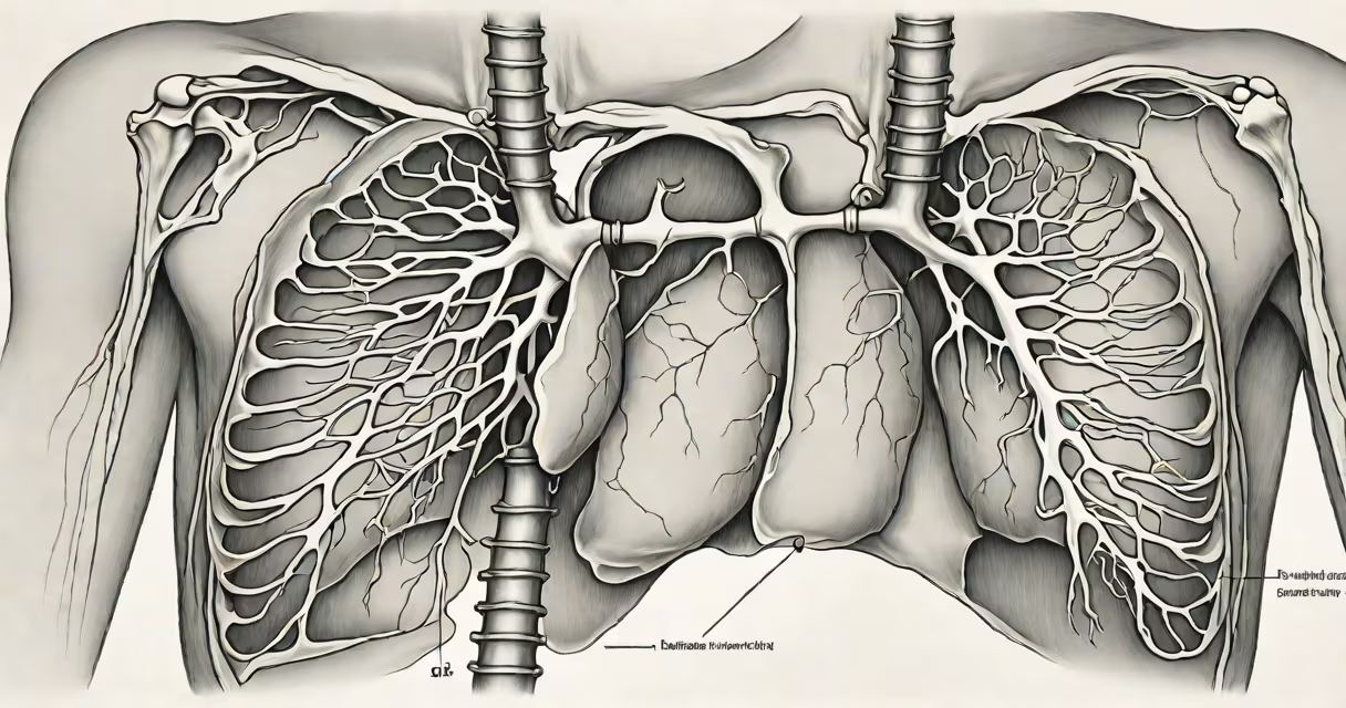
Chest X rays rarely show direct evidence of pulmonary embolism, but specific indirect signs can suggest the condition. These include focal opacities, decreased vascular markings, and pleural effusions-findings that prompt further imaging such as CT pulmonary angiography.
X ray signs of pulmonary embolism are indirect and often subtle. They result from changes in lung perfusion rather than the clot itself. The chest X ray cannot visualize the thrombus, but it detects downstream effects like infarction or altered blood flow. A normal chest X ray does not rule out pulmonary embolism-up to 30% of patients with confirmed PE have a normal initial film.
When abnormalities appear, they typically reflect lung tissue damage due to blocked blood flow. The most recognized signs include the Westermark sign (localized oligemia or reduced vascular markings) and Hampton's hump (a wedge-shaped opacity at the lung periphery). These findings occur in about 10-15% of cases and are more common in large or central clots.
X rays use ionizing radiation to project a two-dimensional image of internal structures. In pulmonary embolism, the clot obstructs pulmonary arteries, reducing blood flow to parts of the lung. This leads to hypoperfusion, which appears on X ray as diminished vascular markings in the affected area. The lack of contrast between vessels and lung tissue creates a "dark" zone compared to surrounding areas.
Infarction-the death of lung tissue due to ischemia-can cause localized consolidation. On X ray, this shows as a wedge-shaped opacity pointing toward the hilum. This is known as Hampton's hump. It develops within 24-48 hours of the event and may resolve over days to weeks. Its presence increases suspicion for PE, especially when combined with clinical risk factors.
The Westermark sign is the opposite: a region of decreased vascularity adjacent to a normal or hyperemic area. It results from arterial occlusion, leading to reduced blood flow and fewer visible vessels. This sign is less sensitive than Hampton's hump but highly specific when seen.
These signs are not diagnostic on their own but serve as red flags. They are most useful when combined with clinical probability scores like Wells or Geneva, and when followed by confirmatory imaging.
| Sign | Description | Sensitivity | Specificity |
|---|---|---|---|
| Hampton's hump | Wedge-shaped opacity near the pleura, base toward the hilum | ~10% | High |
| Westermark sign | Localized absence of pulmonary vessels (oligemia) | ~5% | Very high |
| Pleural effusion | Fluid accumulation in the pleural space | ~20% | Moderate |
| Enlarged pulmonary artery | Visible widening of the main pulmonary artery | ~15% | High |
According to the American College of Radiology (ACR), neither Hampton's hump nor Westermark sign is sufficient for diagnosing pulmonary embolism. However, their presence increases the pre-test probability and warrants urgent CT pulmonary angiography. A study published in *Radiographics* found that when both signs were present, the likelihood of PE exceeded 80%.
One non-obvious fact: Hampton's hump may mimic pneumonia on X ray. Both conditions produce similar opacities. Differentiation relies on clinical context-fever, leukocytosis, and sputum production favor infection. In PE, symptoms are more likely to include sudden dyspnea, pleuritic chest pain, and unexplained tachycardia.
Many clinicians assume that a normal chest X ray rules out pulmonary embolism. This is incorrect. A normal film does not exclude PE. In fact, a 2021 retrospective analysis of over 1,200 emergency department patients showed that 34% of those later diagnosed with PE had normal initial X rays.
Another misconception is that all pulmonary emboli cause visible signs. Large clots in the main or lobar arteries are more likely to produce detectable changes. Small segmental or subsegmental clots often go undetected on X ray. This is why CT pulmonary angiography remains the gold standard for confirmation.
I once reviewed a case where a 62-year-old woman presented with acute shortness of breath. Her chest X ray showed a small right-sided pleural effusion and mild increased lucency in the right lower lobe. The radiologist noted subtle oligemia. The patient was initially misdiagnosed with pneumonia. Only after a CT scan revealed a saddle embolus in the main pulmonary artery did the correct diagnosis emerge. This case highlights how reliance on X ray alone can delay treatment.
Chest X rays are routinely performed in patients with suspected pulmonary embolism as part of the initial workup. They help assess for alternative diagnoses-such as pneumonia, heart failure, or pneumothorax-that may explain symptoms. They also provide baseline anatomy for comparison if follow-up imaging is needed.
If X ray signs of pulmonary embolism are present, the next step is always CT pulmonary angiography. This test visualizes the pulmonary arteries and confirms the presence and location of clots. The sensitivity of CT PA exceeds 95%, making it the definitive tool for diagnosis.
For patients with normal X rays but high clinical suspicion, D-dimer testing is used. A negative D-dimer in low-risk patients effectively rules out PE. In moderate-to-high risk individuals, CT PA is still required even if the X ray is normal.
Understanding x ray signs of pulmonary embolism is essential for frontline clinicians. While these signs are not definitive, recognizing them can trigger timely investigation and reduce delays in treatment. Early intervention lowers mortality rates -untreated PE has a 30-day fatality rate of up to 15%.