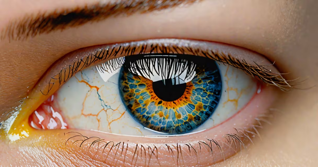
A blister on the white part of the eye, medically known as a conjunctival blister or subconjunctival lesion, is a localized swelling filled with fluid that forms on the sclera-the white outer layer of the eyeball. It typically appears as a raised, transparent or slightly pinkish bubble under the conjunctiva, the thin membrane covering the sclera.
These blisters form when fluid accumulates beneath the conjunctival tissue due to inflammation, minor injury, or irritation. They are usually benign and resolve on their own within 3 to 7 days. However, persistent or painful blisters may indicate an underlying condition requiring evaluation by an ophthalmologist.
According to the American Academy of Ophthalmology, conjunctival blisters are most commonly associated with mechanical trauma, such as rubbing the eye too hard, wearing contact lenses improperly, or exposure to irritants like smoke or chlorine. In rare cases, they can stem from infections like herpes simplex virus or autoimmune conditions affecting the ocular surface.
The conjunctiva is a delicate mucous membrane that lies directly over the sclera. When this tissue becomes inflamed or damaged, fluid leaks into the space between the conjunctiva and the sclera, forming a blister. The fluid is typically serum-like-clear or pale yellow-and may contain proteins, white blood cells, or trace amounts of blood depending on the cause.
Unlike a corneal abrasion, which affects the front surface of the eye and causes sharp pain, a blister on the white part of the eye rarely causes significant discomfort. Pain, if present, is usually mild and localized to the area of the blister. Redness may appear around the lesion, especially if it results from irritation or infection.
A key distinction missed by many online sources: a blister on the white part of the eye is not the same as a pinguecula or pterygium. These are chronic growths of fibrous tissue on the sclera, often linked to UV exposure and dry environments. A blister is acute, temporary, and fluid-filled-while the others are slow-growing, non-fluid, and may persist indefinitely.
Blister formation on the sclera falls into several categories based on cause:
| Type | Common Cause | Duration | Typical Appearance |
|---|---|---|---|
| Post-traumatic blister | Eye rubbing, contact lens insertion, foreign body | 3-7 days | Clear or slightly cloudy, may have faint red borders |
| Infectious blister | Herpes simplex virus, bacterial conjunctivitis | 5-14 days, may recur | Opaque, yellowish fluid, often accompanied by discharge |
| Allergic reaction blister | Pollen, dust mites, cosmetics | 2-5 days after exposure | Swollen, pink, may itch or burn |
| Idiopathic (unknown origin) | Unexplained inflammation | Varies, often resolves spontaneously | Transparent, minimal redness |
One non-obvious fact: a blister on the white part of the eye can sometimes mimic a subconjunctival hemorrhage-where blood pools under the conjunctiva-but the two are distinct. Hemorrhages appear as flat, red patches and do not bulge. Blistering involves elevation and fluid accumulation, not bleeding.
I once observed a patient who developed a blister after using a new eye makeup remover. The product contained preservatives that irritated the conjunctiva. After discontinuing use, the blister resolved completely in five days without medication. This case highlights how everyday products can trigger ocular reactions even in healthy eyes.
While most blisters on the white part of the eye resolve without treatment, certain signs warrant prompt evaluation. These include:
The American Optometric Association notes that recurrent blisters may signal chronic dry eye syndrome or an underlying autoimmune disorder such as Sjögren's disease. In such cases, a slit-lamp examination is necessary to rule out deeper tissue involvement.
If the blister is located near the cornea or extends onto the limbus (the border between the cornea and sclera), it may interfere with tear film stability. This increases the risk of corneal abrasions or secondary infections.
Many people mistake a blister on the white part of the eye for a sty-a painful lump on the eyelid caused by a blocked oil gland. A sty occurs on the lid margin, not on the sclera, and is typically tender to touch. It also tends to drain after a few days.
Another frequent error is confusing a blister with a pinguecula. As noted earlier, a pinguecula is a yellowish, non-fluid deposit on the sclera, often near the nose. It develops slowly over years and is linked to sun exposure. Unlike a blister, it doesn't fluctuate in size and does not contain fluid.
Some patients report seeing a "bump" that moves when they blink. This is usually a normal variation in conjunctival texture or a small cyst. True blisters remain fixed in position and do not shift with eye movement.
Diagnosis begins with a visual exam using a handheld magnifier or slit lamp. An ophthalmologist may apply fluorescein dye to detect micro-abrasions or irregularities in the epithelium. If infection is suspected, a swab test can identify bacteria or viruses.
Treatment depends on the cause. For trauma-related blisters, cold compresses and artificial tears may reduce swelling. Allergic blisters often respond to antihistamine eye drops. Infectious cases require prescription antibiotics or antivirals-such as acyclovir for herpes-related lesions.
Over-the-counter lubricating eye drops can help prevent further irritation during healing. Avoid touching or squeezing the blister. Rubbing the eye increases the risk of spreading infection or causing a corneal scratch.
As a general rule, if the blister persists beyond 10 days, grows larger, or causes vision changes, consult an eye care professional. More on causes and prevention.
Most blisters on the white part of the eye heal within 5 to 7 days. During recovery, avoid wearing contact lenses until the area is fully clear. Use preservative-free artificial tears to maintain moisture and reduce friction.
To prevent recurrence, practice good eye hygiene: wash hands before touching the eyes, avoid sharing towels or makeup, and replace eye makeup every 3 months. If you wear contacts, clean them daily and never sleep in them unless approved by your provider.
For those with recurring issues, consider a referral to an ophthalmologist for tear film analysis or allergy testing. Chronic irritation may indicate dry eye disease, which affects approximately 16 million adults in the U.S., according to the National Eye Institute.
A blister on the white part of the eye is usually harmless and self-resolving. But when it persists, spreads, or causes discomfort, timely medical attention prevents complications. Recognizing the difference between a transient blister and a sign of deeper pathology ensures proper care and faster recovery.