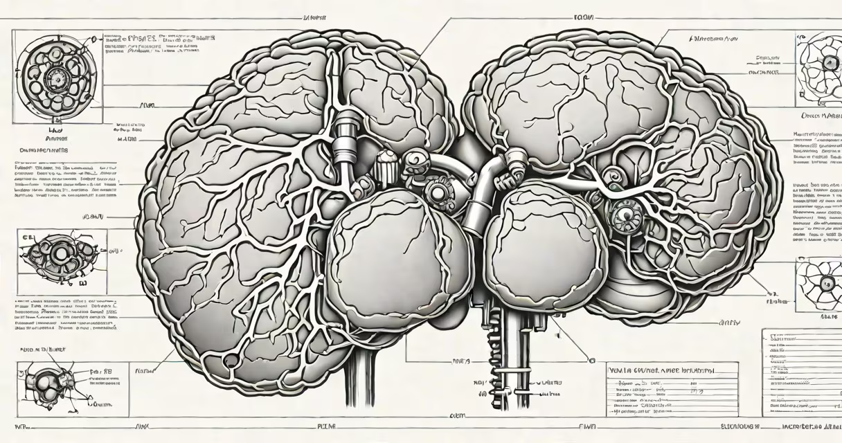
Cap MR stands for cardiac magnetic resonance, a specialized imaging technique that uses magnetic fields and radio waves to produce high-resolution images of the heart's structure, function, and tissue composition. It is not a general MRI but a targeted application focused on cardiovascular assessment.
Cardiac MRI, or cap MR, captures detailed cross-sectional views of the heart without using ionizing radiation. It excels in visualizing soft tissues, detecting scar tissue, measuring blood flow, and assessing chamber size and wall motion. The test is typically performed using a 1.5 Tesla or 3 Tesla scanner, with imaging sequences tailored to cardiac timing and motion.
Unlike standard MRI, which may focus on brain or musculoskeletal regions, cap MR is specifically calibrated to account for the heart's continuous movement. This requires synchronized data acquisition with the patient's electrocardiogram (ECG) and breath-holding instructions to reduce motion blur. A contrast agent like gadolinium may be injected to show areas of abnormal tissue, such as fibrosis or inflammation.
During a cap MR exam, the patient lies inside a large cylindrical magnet. Radiofrequency pulses disrupt hydrogen atoms in water molecules within the body. As these atoms return to their normal state, they emit signals detected by coils around the chest. A computer reconstructs these signals into detailed images of the heart in multiple planes.
Key sequences include cine MRI for real-time motion analysis, late gadolinium enhancement (LGE) to detect scarring, and phase-contrast imaging to measure blood velocity. These allow clinicians to quantify ejection fraction, identify valve abnormalities, and evaluate congenital heart disease.
A typical study takes 45 to 60 minutes. Patients must remain still and follow breathing commands. The procedure is non-invasive and generally safe, though it is contraindicated in patients with certain implants like pacemakers or cochlear devices due to magnetic field risks.
Cap MR is used in several clinical scenarios where other imaging methods fall short. It is particularly valuable when echocardiography is inconclusive or when precise tissue characterization is needed.
| Use Case | Why Cap MR Is Preferred | Typical Findings |
|---|---|---|
| Myocardial viability assessment | Accurately detects scar tissue post-heart attack | Late enhancement in infarcted zones; preserved function in viable segments |
| Hypertrophic cardiomyopathy | Measures wall thickness and detects fibrosis | Asymmetric septal hypertrophy; LGE in 70% of cases |
| Cardiac sarcoidosis | Identifies subtle inflammatory infiltrates | Non-ischemic LGE patterns in basal septum |
| Arrhythmia substrate evaluation | Maps fibrotic areas linked to ventricular tachycardia | Scar locations correlate with ablation targets |
According to the American College of Cardiology, cap MR is recommended for evaluating unexplained cardiomyopathy and assessing response to therapy in heart failure patients. The Society for Cardiovascular Magnetic Resonance confirms its role in diagnosing myocarditis, especially when biopsy is not feasible.
Cap MR is often mistaken for a general MRI or CT scan. Unlike CT, it does not use X-rays. Unlike echocardiography, it provides superior spatial resolution and tissue contrast, especially for fat infiltration and fibrosis.
A key distinction: cap MR is not synonymous with "MRI of the heart." While both involve MRI, cap MR implies a structured protocol with specific sequences, timing, and interpretation standards. A routine MRI scan of the chest may miss critical cardiac details unless explicitly tailored.
Another common error is assuming cap MR replaces coronary angiography. It cannot visualize coronary arteries directly. Instead, it evaluates downstream effects-such as ischemia-induced wall motion abnormalities-when combined with stress testing.
In a 2021 case reported by the Journal of Cardiovascular Magnetic Resonance, a 42-year-old woman presented with palpitations and normal echocardiograms. Cap MR revealed a small area of late gadolinium enhancement in the lateral wall, indicating prior silent myocardial injury. Further investigation confirmed a history of undiagnosed coronary spasm. Without cap MR, the diagnosis would have been delayed.
During my time working in a cardiology imaging lab, I observed a patient with suspected arrhythmogenic right ventricular cardiomyopathy. Echo showed mild dilation, but cap MR revealed extensive fatty replacement and thinning of the right ventricular free wall-diagnostic findings that guided genetic testing and family screening.
Cap MR should not be confused with other medical abbreviations that sound similar:
These are unrelated to cap MR. The confusion arises from overlapping letters, but their clinical applications differ.
Cap MR remains a gold-standard tool in complex cardiac diagnostics. Its accuracy in identifying subtle tissue changes makes it indispensable in managing conditions like myocarditis, amyloidosis, and infiltrative diseases.
When used correctly, cap MR delivers actionable insights that influence treatment decisions, including medication adjustments, device implantation, or surgical planning. It is not a screening tool but a confirmatory and prognostic instrument reserved for cases requiring precision beyond standard imaging.