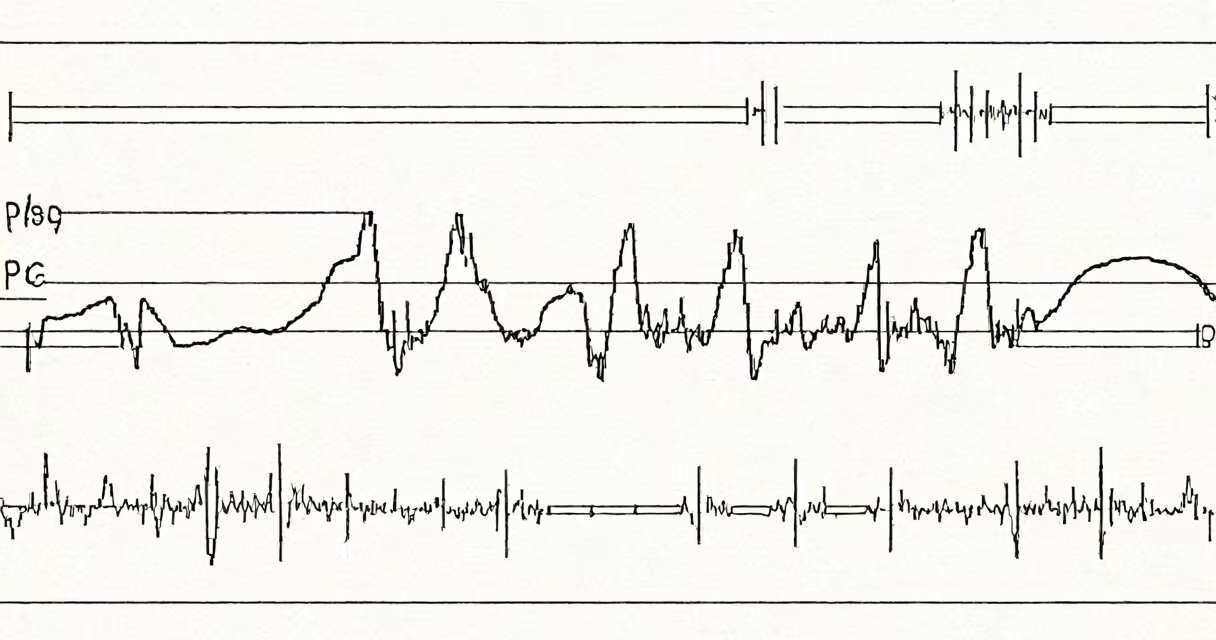
Supraventricular tachycardia (SVT) ECG interpretation identifies a rapid heart rhythm originating above the ventricles, typically with a rate exceeding 150 beats per minute, narrow QRS complexes (<120 ms), and often absent or buried P waves. The rhythm appears regular and can mimic sinus tachycardia, but the absence of P waves and sudden onset distinguish it clinically.
SVT arises from abnormal electrical circuits in the atria or the AV node. The most common form is AV nodal reentrant tachycardia (AVNRT), which accounts for about 60% of cases. Other types include AV reentrant tachycardia (AVRT), often associated with Wolff-Parkinson-White syndrome, and focal atrial tachycardia. ECG interpretation focuses on distinguishing these based on morphology, timing, and response to maneuvers.
When interpreting an ECG during SVT, look for a regular rhythm, narrow QRS complexes, and a rate between 150 and 250 bpm. P waves may be absent, inverted, or hidden within the QRS complex. The absence of discernible P waves is a sign of AVNRT. In contrast, AVRT may show retrograde P waves immediately following the QRS, creating a "pseudo" negative deflection in leads II, III, and aVF.
Narrow-complex tachycardias are categorized by QRS width. A QRS duration under 120 ms indicates supraventricular origin. Wide-complex tachycardias (>120 ms) suggest ventricular origin or aberrant conduction. Misinterpreting SVT as ventricular tachycardia (VT) can lead to inappropriate treatment, such as administering adenosine to a VT patient, which risks worsening hemodynamics.
Key differentiators include the presence of AV dissociation (common in VT), capture beats, fusion beats, and a history of structural heart disease. In SVT, AV dissociation is absent. Instead, AV conduction remains intact, resulting in a 1:1 relationship between P waves and QRS complexes - though P waves may be obscured.
A 2021 study in Circulation: Arrhythmia and Electrophysiology found that 87% of SVT episodes in emergency settings were misdiagnosed initially due to overlapping features with sinus tachycardia or atrial fibrillation with rapid ventricular response. This underscores the need for precise ECG analysis rather than relying solely on rate or symptoms.
The classic ECG pattern of AVNRT shows a regular narrow-complex tachycardia with no visible P waves. Instead, a small negative deflection may appear just after the QRS in inferior leads, indicating retrograde activation of the atria. This is known as a "pseudo-negative" P wave.
For AVRT, the ECG often displays a delta wave in the baseline tracing if WPW is present. During SVT, the delta wave disappears during the tachycardia, and the QRS becomes narrow. Retrograde P waves are frequently seen immediately after the QRS, especially in leads II, III, and aVF.
| Type of SVT | Typical Rate (bpm) | QRS Width | P Wave Appearance | Common Triggers |
|---|---|---|---|---|
| AV nodal reentrant tachycardia (AVNRT) | 150-250 | <120 ms | Often absent or buried | Stress, caffeine, alcohol |
| AV reentrant tachycardia (AVRT) | 150-250 | <120 ms | Retrograde P wave after QRS | WPW syndrome, congenital accessory pathways |
| Focal atrial tachycardia | 100-150 | <120 ms | Visible, upright P waves in inferior leads | Hyperthyroidism, electrolyte imbalance |
One non-obvious distinction: in AVNRT, the retrograde P wave is usually not visible because the atria depolarize after the ventricles. In AVRT, the retrograde P wave is often visible due to the longer refractory period in the accessory pathway. This subtle difference aids in differentiation when the rhythm is unclear.
In a 28-year-old woman presenting with palpitations and lightheadedness, the ECG showed a regular narrow-complex tachycardia at 180 bpm. QRS complexes were 96 ms wide. No distinct P waves were visible. There was a small downward deflection immediately following each QRS in leads II, III, and aVF.
These findings met criteria for AVNRT. Adenosine was administered intravenously, resulting in abrupt termination of the rhythm and return to normal sinus rhythm. The diagnosis was confirmed via electrophysiological study later, which demonstrated dual AV nodal pathways.
During my time as a cardiology fellow, I encountered a similar case where a patient with a history of panic attacks had a prolonged episode of SVT. The initial ECG was misread as sinus tachycardia due to the high rate. Only after performing a carotid massage and observing the sudden cessation of tachycardia did we confirm the diagnosis. This experience reinforced that ECG interpretation must go beyond rate and consider rhythm regularity and P wave visibility.
One frequent error is confusing SVT with atrial fibrillation with rapid ventricular response. In AFib, the rhythm is irregularly irregular, and P waves are replaced by fibrillatory waves. In SVT, the rhythm is regular. Atrial flutter with variable conduction can mimic SVT, but it shows sawtooth flutter waves in inferior leads, particularly in lead II.
Another mistake is assuming all narrow-complex tachycardias are SVT. Some patients with structurally normal hearts develop idiopathic ventricular tachycardia, which can appear narrow if the focus is near the AV node. These cases require careful evaluation, including a history of syncope or family history of sudden death.
According to the American Heart Association's 2023 guidelines on acute management of tachyarrhythmias, adenosine remains the first-line agent for stable SVT. Its mechanism involves transiently blocking AV nodal conduction, allowing the intrinsic pacemaker to resume control. Response occurs within 30 seconds in 90% of AVNRT cases.
For unstable patients, synchronized cardioversion at 50-100 J is recommended. However, this is rarely needed in SVT due to the high tolerance of the rhythm in otherwise healthy individuals.
Understanding supraventricular tachycardia ECG interpretation enables accurate diagnosis and timely intervention. When in doubt, compare the ECG to a baseline rhythm strip and assess for P wave presence, QRS width, and rhythm regularity. Always correlate findings with the patient's clinical status.