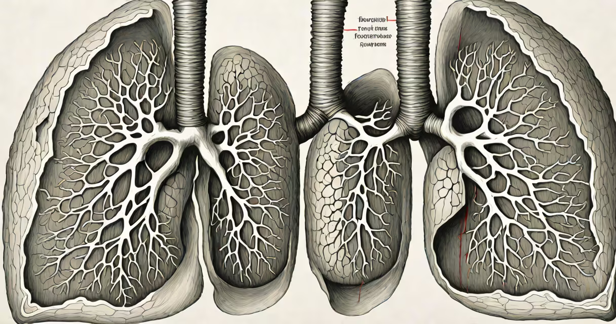
ventilation perfusion mismatch, or V/Q mismatch, occurs when the amount of air reaching the alveoli (ventilation) doesn't match the amount of blood flowing through the pulmonary capillaries (perfusion), disrupting efficient oxygen and carbon dioxide exchange in the lungs.
This imbalance reduces arterial oxygen saturation and can lead to hypoxemia-low oxygen in the blood-even when breathing room air. It's a core mechanism behind many respiratory conditions, including pneumonia, pulmonary embolism, and chronic obstructive pulmonary disease (COPD).
The lungs maintain optimal gas exchange through precise matching of ventilation and perfusion. When this balance fails, areas of the lung either receive air without adequate blood flow (high V/Q ratio) or blood without sufficient air (low V/Q ratio). Both scenarios impair oxygen uptake and carbon dioxide removal.
Each alveolus requires both air and blood to function. Air enters via the bronchial tree and reaches alveoli during inhalation. Simultaneously, deoxygenated blood arrives from the right side of the heart through pulmonary arteries. Oxygen diffuses from alveoli into red blood cells, while carbon dioxide moves in the opposite direction.
In healthy lungs, ventilation and perfusion are well-matched. The ideal V/Q ratio is approximately 0.8-meaning for every liter of air entering the alveoli, about 1.25 liters of blood flow through the capillaries. This ratio ensures maximum efficiency in gas exchange.
When mismatched, gas exchange becomes inefficient. For example, if a region of the lung is ventilated but not perfused (e.g., due to a pulmonary embolism), the air reaches alveoli but no blood is available to pick up oxygen. This area acts as dead space, wasting ventilation.
V/Q mismatch is categorized by the degree of imbalance between ventilation and perfusion. The most common types are:
| Type | Definition | Common Causes | Effect on Blood Gases |
|---|---|---|---|
| Low V/Q | Perfusion exceeds ventilation | Pneumonia, atelectasis, asthma | Arterial hypoxemia, normal or low PaCO₂ |
| High V/Q | Ventilation exceeds perfusion | Pulmonary embolism, reduced cardiac output | Increased alveolar dead space, normal PaO₂ unless severe |
| Shunt | No ventilation despite perfusion | Consolidated lung tissue, fluid-filled alveoli | Severe hypoxemia unresponsive to supplemental oxygen |
Low V/Q is the most frequent cause of hypoxemia in clinical settings. High V/Q contributes less to oxygen loss but increases dead space. Shunts are particularly dangerous because they bypass gas exchange entirely.
A key insight often missed: shunts do not respond to increased FiO₂ (fraction of inspired oxygen), unlike low V/Q regions. This distinction is critical in diagnosing and managing respiratory failure.
V/Q mismatch underlies nearly all forms of acute and chronic respiratory dysfunction. In pneumonia, inflammatory exudate fills alveoli, preventing ventilation even though blood continues to flow-this creates a shunt. In pulmonary embolism, clots block pulmonary vessels, reducing perfusion to ventilated areas-resulting in high V/Q.
COPD patients develop regional V/Q mismatch due to airway obstruction and destruction of alveolar walls. In emphysema, alveoli lose elasticity and collapse during expiration, trapping air and creating hyperinflated zones with poor perfusion. In chronic bronchitis, mucus plugs obstruct airflow, leading to poorly ventilated but still perfused regions.
Even in healthy individuals, gravity causes a natural gradient: apical lung regions receive more ventilation relative to perfusion (V/Q ~3.0), while basal regions have better matching (V/Q ~0.6). This physiological variation is normal and usually compensated by hypoxic vasoconstriction in poorly ventilated areas.
Many assume V/Q mismatch only affects people with lung disease. But it occurs during anesthesia, in critically ill patients, and even in healthy people lying supine. A 2021 study using lung imaging showed that up to 40% of healthy adults exhibit measurable V/Q heterogeneity during sleep.
Another misconception: that all hypoxemia stems from V/Q mismatch. While it's a major contributor, other mechanisms exist-such as diffusion impairment (e.g., pulmonary fibrosis), hypoventilation (e.g., opioid overdose), and right-to-left shunting (e.g., congenital heart defects).
V/Q mismatch does not always cause elevated CO₂. In early stages, patients may compensate by increasing minute ventilation. Only when the mismatch is severe or compensation fails does hypercapnia develop.
Consider a 62-year-old woman presenting with sudden shortness of breath and chest pain. Arterial blood gas shows PaO₂ = 60 mmHg (normal ~80-100), PaCO₂ = 35 mmHg (normal), and pH = 7.45. Her oxygen saturation drops to 88% on room air.
A CT pulmonary angiogram reveals a clot blocking the left pulmonary artery. This reduces perfusion to the left lower lobe while ventilation remains intact-creating a high V/Q state. The alveoli fill with air but lack blood flow, so oxygen cannot transfer into circulation.
Her hypoxemia results from wasted ventilation. Supplemental oxygen improves saturation slightly but not completely-because the problem lies in perfusion, not oxygen availability. Treatment involves anticoagulation and, in severe cases, thrombolysis.
I once observed a patient in the ICU with sepsis-induced ARDS. Despite receiving 60% oxygen, their SpO₂ remained below 90%. A V/Q scan revealed widespread low V/Q regions due to alveolar flooding from inflammation. The patient required prone positioning and PEEP (positive end-expiratory pressure) to recruit collapsed alveoli and improve ventilation-perfusion alignment.
That case underscored a key point: correcting V/Q mismatch isn't always about giving more oxygen. Sometimes, improving lung mechanics or recruiting collapsed units matters more.
Understanding ventilation perfusion mismatch allows clinicians to differentiate between causes of hypoxemia and choose appropriate interventions. It explains why some patients remain hypoxic despite high-flow oxygen and why certain treatments-like PEEP, recruitment maneuvers, or anticoagulants-are effective.
For patients, recognizing symptoms like unexplained shortness of breath, especially with exertion, can prompt earlier evaluation. Early detection prevents progression to respiratory failure.
Ventilation perfusion mismatch is not a single diagnosis-it's a pathophysiological process underlying many respiratory disorders. Recognizing it enables targeted therapy, better outcomes, and reduced reliance on invasive support.