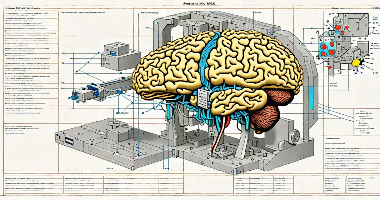
An fMRI, or functional magnetic resonance imaging, is a non-invasive medical imaging technique that measures brain activity by detecting changes in blood flow. It shows which areas of the brain are engaged during specific tasks or states, such as thinking, remembering, or feeling emotions.
fMRI scans do not measure neural firing directly. Instead, they rely on the blood-oxygen-level-dependent (BOLD) signal-a proxy for neuronal activity. When neurons fire, they consume oxygen, and nearby blood vessels dilate to deliver more oxygen-rich blood. The fMRI detects the increased concentration of oxygenated hemoglobin in those areas, indicating heightened activity.
The technique was first demonstrated in 1990 by researchers at the University of California, San Diego, and has since become a key part of cognitive neuroscience. It allows scientists to observe brain function in real time, with spatial resolution down to about 1-3 millimeters and temporal resolution around 1-2 seconds per image slice.
fMRI uses a useful magnet to align hydrogen atoms in water molecules within the brain. Radio waves are then pulsed through the head, causing the atoms to emit signals as they return to alignment. These signals are captured by sensors and converted into detailed images of brain structure and function.
During a scan, participants lie still inside a cylindrical scanner while performing tasks-such as viewing images, solving math problems, or listening to sounds. The scanner captures hundreds of images over several minutes, comparing differences in blood flow between baseline states and task periods.
A key advantage of fMRI is its ability to localize activity across the entire brain simultaneously. Unlike EEG, which tracks electrical activity with high temporal precision but poor spatial accuracy, fMRI offers both good spatial detail and reasonable timing resolution. However, it cannot detect events occurring faster than the BOLD response, which typically peaks 4-6 seconds after neural activation.
There are two primary forms of fMRI based on the type of data collected:
| Type | Measures | Use Case Example | Limitation |
|---|---|---|---|
| BOLD fMRI | Changes in oxygenated blood flow | Mapping language centers in patients before brain surgery | Indirect measure; delayed response |
| Arterial spin labeling (ASL) | Direct measurement of cerebral blood flow | Studying brain perfusion in stroke recovery | Lower signal-to-noise ratio; longer scan times |
BOLD fMRI remains the most widely used method due to its compatibility with standard MRI hardware and ease of implementation. ASL is less common but valuable in clinical settings where direct blood flow measurements are needed, such as monitoring patients with vascular dementia or traumatic brain injury.
Functional MRI is also used in resting-state fMRI, where participants lie quietly without performing tasks. Researchers analyze spontaneous fluctuations in the BOLD signal to identify intrinsic brain networks-like the default mode network, which activates during mind-wandering and self-referential thought.
Many people assume fMRI shows "thoughts" directly. This is incorrect. The scan reveals patterns of blood flow associated with neural processing, not conscious content. You cannot read someone's thoughts from an fMRI image alone.
Another misconception is that fMRI can detect lies. While certain brain regions show different activity during truthful versus deceptive responses, no fMRI-based lie detection system is legally admissible or scientifically validated for forensic use. The American Psychological Association has stated that fMRI cannot reliably distinguish truth from deception under real-world conditions.
A lesser-known fact: fMRI data often requires complex statistical modeling to interpret. Raw images appear as noisy color maps. Researchers apply algorithms like general linear modeling (GLM) to isolate meaningful signals from background noise. A single study may involve tens of thousands of data points analyzed across multiple brain regions.
In clinical practice, fMRI helps surgeons avoid damaging critical brain areas during tumor removal. For example, a patient with a glioma near Broca's area-an area responsible for speech production-might undergo a preoperative fMRI to map language function. If the scan shows that speech-related activity overlaps with the tumor, the surgical team adjusts their approach to preserve function.
In research, fMRI has identified brain regions linked to depression. A 2018 study published in Nature Neuroscience found that individuals with major depressive disorder showed reduced connectivity in the prefrontal cortex and increased activity in the amygdala during emotional processing tasks. These findings support the development of targeted treatments, including transcranial magnetic stimulation (TMS).
I once observed an fMRI session at a university lab where a participant was asked to imagine walking through a familiar city street. The scan showed consistent activation in the hippocampus and posterior cingulate cortex-areas involved in spatial memory and autobiographical recall. The pattern matched known neural signatures of mental imagery, confirming that fMRI can capture internal experiences even without physical movement.
fMRI is not a tool for reading minds or diagnosing mental illness on its own. It is a research and diagnostic aid that provides insights into how the brain functions during specific behaviors or states. Its strength lies in correlating brain activity with behavior, not in predicting individual thoughts or decisions.
While fMRI has limitations-including cost ($1,000-$3,000 per scan), long acquisition times (15-45 minutes), and sensitivity to motion-the benefits far outweigh the drawbacks in controlled environments. It continues to advance our understanding of the human brain, from basic cognition to complex disorders like Alzheimer's and schizophrenia.
Understanding what is an fMRI begins with recognizing it as a window-not a mirror. It shows where activity occurs, not why it happens. Interpreting results requires careful experimental design, statistical rigor, and integration with other data sources.
For deeper insight into brain function, fMRI is unmatched. But it must be used correctly, interpreted cautiously, and never treated as definitive proof of internal states.