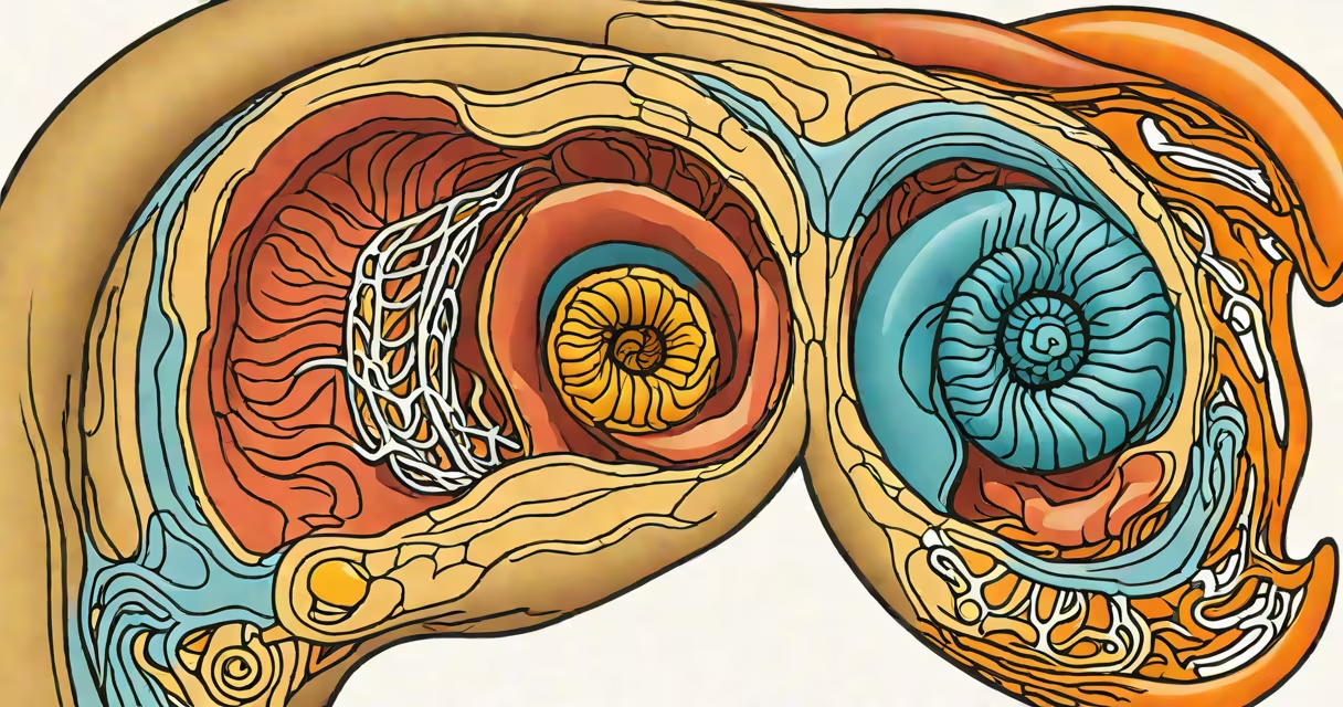
The inner ear is formally known as the labyrinth, a network of fluid-filled structures within the temporal bone that govern hearing and balance. It consists of the cochlea for sound perception and the vestibular system for spatial orientation.
The term "labyrinth" reflects its complex layout-named for its maze-like structure. This system includes the cochlea, which converts sound waves into neural signals, and the vestibular apparatus, composed of three semicircular canals and two otolith organs (utricle and saccule). These components detect motion and head position, enabling stable posture and coordination during movement.
According to the National Institute on Deafness and Other Communication Disorders (NIDCD), the inner ear's sensory cells are highly sensitive and easily damaged by loud noise, aging, or certain medications. Once damaged, they do not regenerate in humans, making prevention critical.
Sound enters the outer ear and travels through the ear canal to the eardrum. Vibrations pass through the middle ear bones-malleus, incus, and stapes-and reach the fluid-filled cochlea. Inside the cochlea, hair cells bend in response to pressure waves, triggering electrical impulses sent via the auditory nerve to the brain.
For balance, the semicircular canals detect rotational motion. Each canal contains endolymph fluid and a cupula-a gelatinous structure that deflects when the head turns. The deflection stimulates hair cells, signaling angular acceleration. The otolith organs respond to linear motion and gravity. Otoliths (tiny calcium carbonate crystals) rest on a gel layer above hair cells; when the head tilts, the crystals shift, bending the hairs and sending positional data.
A real-world example: When you spin around quickly and stop, your head stops moving, but the fluid in the semicircular canals keeps swirling. This mismatch causes dizziness until the fluid settles-this is why spinning can make you feel nauseous.
The inner ear comprises three main parts: the cochlea, the vestibule, and the semicircular canals. All are housed within the bony labyrinth, a rigid casing that protects delicate internal structures.
| Structure | Function | Related Condition |
|---|---|---|
| Cochlea | Converts sound vibrations into neural signals | Presbycusis (age-related hearing loss) |
| Semicircular canals | Detect rotation and head tilt | Benign paroxysmal positional vertigo (BPPV) |
| Utricle and saccule | Respond to linear acceleration and gravity | Otoconia dislodgement (BPPV) |
Each semicircular canal lies in a different plane-horizontal, anterior, and posterior-to monitor movement in all directions. The utricle senses forward-backward and side-to-side motion, while the saccule detects vertical movement and static head position relative to gravity.
Damage to any component disrupts sensory input. For instance, Meniere's disease involves excess fluid in the cochlea and vestibular system, causing episodes of vertigo, tinnitus, and fluctuating hearing loss. The American Academy of Otolaryngology-Head and Neck Surgery notes that about 1 in 1,000 people develop Meniere's disease annually.
Many assume the inner ear is simply a passive receiver of sound. In reality, it actively processes and filters auditory input. The cochlea performs frequency analysis-different regions respond to specific pitches-before transmitting data to the brainstem and auditory cortex.
Another misconception is that balance issues always stem from inner ear problems. While the inner ear plays a central role, neurological conditions such as multiple sclerosis or stroke can mimic vestibular symptoms. Distinguishing between peripheral (inner ear) and central (brain) causes requires clinical evaluation.
A lesser-known fact: The inner ear shares embryonic origin with the eye. Both develop from the ectoderm layer during early fetal development. This explains why some genetic disorders affect both vision and hearing-such as Usher syndrome, which causes progressive deafness and retinitis pigmentosa.
I once observed a patient with BPPV during a clinic visit. After lying down and turning their head, they reported sudden vertigo lasting seconds. A Dix-Hallpike maneuver confirmed displaced otoconia in the posterior semicircular canal. Repositioning therapy-performed in under five minutes-resolved the episode. This demonstrated how a tiny crystal displacement can trigger dramatic symptoms.
Inner ear disorders affect quality of life. Vertigo episodes can lead to falls, especially in older adults. The CDC reports that nearly 1 in 3 Americans aged 65 and older experience balance problems annually, with inner ear dysfunction being a leading cause.
Understanding the inner ear as the labyrinth clarifies its dual role in hearing and equilibrium. Its name reflects complexity, not just anatomy. The system's sensitivity to damage underscores the importance of protecting it-through noise reduction, avoiding ototoxic drugs, and seeking prompt care for symptoms like persistent dizziness or hearing changes.
When you ask "what is the inner ear called," the answer is the labyrinth. This structure integrates auditory and vestibular functions through specialized fluid dynamics and microscopic hair cells. It is not merely a hearing organ-it is essential for spatial awareness, stability, and environmental navigation.