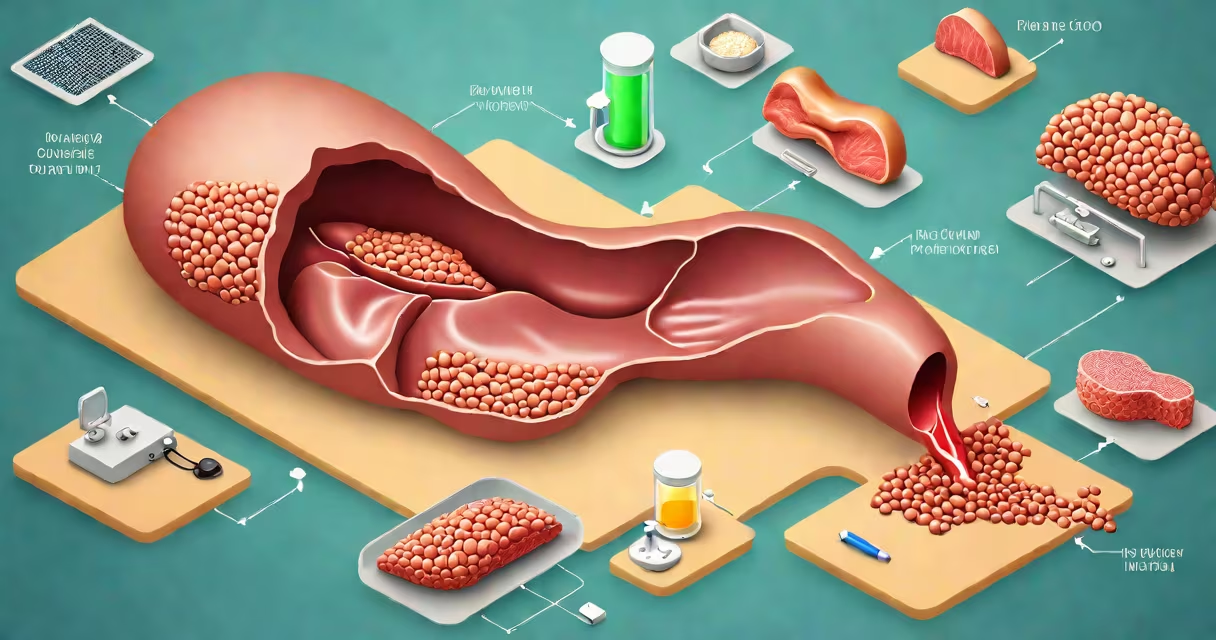
An enlarged liver, or hepatomegaly, occurs when the organ exceeds normal size due to underlying medical conditions. It's not a diagnosis itself but a sign of potential liver dysfunction. The most common causes include fatty liver disease, chronic alcohol use, viral infections like hepatitis B and C, and metabolic disorders such as hemochromatosis.
Doctors detect hepatomegaly during physical exams or imaging tests like ultrasound, CT, or MRI. A liver that's palpable below the rib cage or measures over 15 cm on imaging is considered enlarged. This condition often develops silently-many people have no symptoms until significant damage occurs.
According to the American Liver Foundation, approximately 25% of adults in the U.S. have non-alcoholic fatty liver disease (NAFLD), which is the leading cause of liver enlargement. When fat accumulates in liver cells, it triggers inflammation and fibrosis. Left unchecked, NAFLD can progress to non-alcoholic steatohepatitis (NASH), cirrhosis, and even liver failure.
The liver performs over 500 important functions, including detoxifying blood, processing nutrients, and producing proteins. When stressed by toxins, viruses, or metabolic overload, liver cells swell and scar tissue forms. This leads to increased size and reduced function.
For example, a patient with type 2 diabetes and obesity may develop insulin resistance. This disrupts fat metabolism, causing triglycerides to build up in hepatocytes. Over time, this fat accumulation distorts liver architecture and increases organ volume. Ultrasound scans show a brighter, more echogenic liver-known as "bright liver" in clinical terms.
A 2021 study published in Hepatology found that 38% of patients with NAFLD had elevated liver enzymes (ALT/AST) and visible hepatomegaly on imaging, even without symptoms. This underscores that liver enlargement often precedes clinical warning signs.
Several distinct conditions lead to liver enlargement. The table below outlines key causes, their prevalence, and diagnostic markers.
| Condition | Prevalence in U.S. Adults | Key Mechanism | Diagnostic Clue |
|---|---|---|---|
| Fatty liver disease (NAFLD/NASH) | 25% | Excess fat deposition in liver cells | Increased liver echo texture on ultrasound |
| Alcohol-related liver disease | 10-15% | Chronic ethanol exposure damages hepatocytes | Elevated GGT and AST > ALT ratio |
| Viral hepatitis (B/C) | 1-2% (Hep B), 1.6% (Hep C) | Viruses replicate in liver cells, triggering immune response | Positive HBsAg or anti-HCV antibodies |
| Cirrhosis (from any cause) | 1-2% | Scarring replaces functional tissue | Irregular liver surface on imaging, portal hypertension |
| Metabolic disorders (e.g., hemochromatosis) | 0.3-0.5% | Iron overload damages liver cells | High serum ferritin and transferrin saturation |
Non-alcoholic fatty liver disease accounts for nearly half of all cases of hepatomegaly in primary care settings. It's strongly linked to obesity, insulin resistance, and high triglyceride levels. A 2020 report from the National Institutes of Health noted that NAFLD prevalence doubled between 2000 and 2020, driven by rising rates of metabolic syndrome.
Alcohol-related liver disease remains a major cause, especially among individuals consuming more than 30 grams of ethanol daily (about 2-3 standard drinks). The liver metabolizes alcohol through the enzyme alcohol dehydrogenase, producing acetaldehyde-a toxic byproduct that damages cell membranes and promotes fat accumulation.
Many assume that an enlarged liver always indicates serious illness. That's not true. Temporary enlargement can occur due to acute infections, medication side effects, or even recent heavy meals. However, persistent enlargement requires evaluation.
A common error is confusing hepatomegaly with splenomegaly-the spleen is located near the liver and can also be enlarged. But the spleen lies under the left rib cage, while the liver is on the right. Imaging distinguishes them. Confusing the two leads to misdiagnosis and unnecessary testing.
Another misconception is that all liver enlargement is visible or painful. In reality, most cases are asymptomatic. A 2019 study in JAMA Internal Medicine showed that 62% of patients with fatty liver disease reported no abdominal pain or fatigue. Symptoms typically appear only when fibrosis or cirrhosis develops.
A 48-year-old man presented with fatigue and mild right upper quadrant discomfort. He had a BMI of 34, fasting glucose of 130 mg/dL, and elevated ALT (110 U/L). An ultrasound revealed a diffusely enlarged liver with increased echogenicity-classic signs of fatty liver disease.
He denied alcohol use. Blood tests ruled out viral hepatitis and autoimmune markers. A FibroScan confirmed stage 2 fibrosis. After six months of dietary changes and exercise, his ALT dropped to 45 U/L, and follow-up imaging showed reduced liver size.
This case illustrates that early detection and lifestyle intervention can reverse liver enlargement. The American Association for the Study of Liver Diseases recommends weight loss of 7-10% to improve liver histology in NAFLD patients.
Enlarged liver is not a standalone condition. It signals the need for investigation into root causes. If you have risk factors-obesity, diabetes, heavy alcohol use, or family history of liver disease-screening is essential.
Red flags include jaundice (yellow skin), dark urine, unexplained weight loss, or swelling in legs and abdomen. These suggest advanced disease. Even without symptoms, routine blood work (liver panel) and imaging can catch issues early.
I've seen patients delay care for years, assuming fatigue was just stress. One man with undiagnosed NASH developed cirrhosis and required a transplant. Early action prevents irreversible damage.
What would cause an enlarged liver? The answer lies in identifying the underlying trigger. With proper screening and management, many cases are reversible. Early diagnosis saves lives.