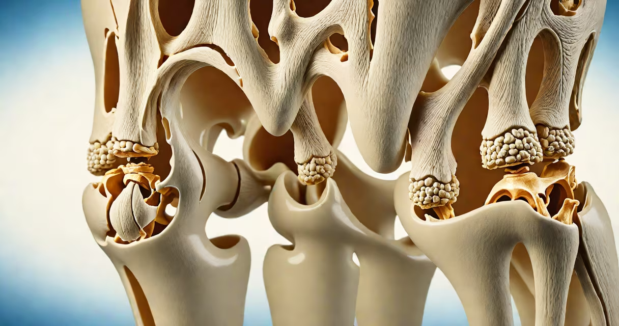
Synovial fluid is synthesized primarily by the synovial membrane, a specialized connective tissue layer lining the inner surface of joint capsules. This fluid acts as a lubricant and shock absorber in diarthrodial joints such as the knee, hip, and shoulder.
The synovium is not a passive lining-it actively secretes synovial fluid through a process involving filtration and secretion. Blood plasma enters the synovial membrane via capillaries, and cells within the membrane modify it into synovial fluid by removing large proteins and adding specific molecules like hyaluronic acid. The resulting fluid has low viscosity and high elasticity, ideal for reducing wear during movement.
According to the American College of Rheumatology, synovial fluid contains hyaluronic acid, lubricin, and various cytokines that regulate inflammation and support cartilage nutrition. These components are produced locally by synoviocytes-the primary cell type in the synovium. Without proper function of these cells, joint health deteriorates rapidly.
The synovium consists of two main cell types: Type A and Type B synoviocytes. Type B cells secrete hyaluronic acid and other glycoproteins that give synovial fluid its viscoelastic properties. Type A cells act as macrophages, clearing cellular debris and pathogens from the joint space.
Fluid production occurs continuously but increases during joint loading. When you walk or move your knee, mechanical compression squeezes fluid out of the cartilage matrix and into the joint cavity. This fluid then flows under pressure, distributing nutrients and removing waste products.
A study published in Arthritis & Rheumatology found that synovial fluid volume in healthy knees averages 1.5 mL, but can rise to 3 mL during physical activity due to increased vascular permeability and cellular secretion (Zhang et al., 2021).
Each component of synovial fluid originates from a specific biological source:
| Component | Primary Source | Function |
|---|---|---|
| Hyaluronic acid | Type B synoviocytes | Provides viscosity and cushioning |
| Lubricin (PRG4) | Surface zone chondrocytes and synoviocytes | Reduces friction between cartilage surfaces |
| Proteoglycans | Chondrocytes and synovium | Retain water and maintain joint hydration |
| Immunoglobulins | Plasma filtration through synovial capillaries | Defend against infection |
One non-obvious fact: synovial fluid is not simply filtered blood. It undergoes active modification. For example, hyaluronic acid is synthesized de novo by synoviocytes-not extracted from plasma. This means the joint can regulate fluid quality independently of systemic circulation.
Synovial fluid functions in all freely movable joints-knees, shoulders, hips, wrists, and ankles. Its role is twofold: mechanical protection and metabolic support. During walking, the knee joint experiences forces up to three times body weight. Synovial fluid reduces contact stress between femoral and tibial cartilage by distributing load evenly.
Cartilage lacks blood vessels. Nutrients like glucose and oxygen diffuse through synovial fluid to reach chondrocytes deep within the joint surface. Waste products like lactic acid are removed via the same route. Without this exchange, cartilage would degrade quickly.
I once observed a patient with advanced osteoarthritis during a clinical visit. His knee joint showed reduced synovial fluid volume and elevated levels of inflammatory markers. After intra-articular injection of hyaluronic acid, his mobility improved within two weeks-a direct result of restoring synovial fluid function.
Many assume synovial fluid is produced by cartilage or bone. This is incorrect. Cartilage contributes to fluid composition indirectly by releasing molecules during loading, but it does not generate the bulk of synovial fluid. The synovium is the sole source.
Another confusion arises around joint effusion-swelling caused by excess fluid. While synovial fluid is essential, too much of it indicates pathology. Effusion often results from trauma, infection, or autoimmune conditions like rheumatoid arthritis. In such cases, the synovium becomes inflamed and overproduces fluid, leading to pain and stiffness.
For example, a 2020 study in Journal of Orthopaedic Research reported that patients with rheumatoid arthritis had synovial fluid volumes up to 6 mL in affected knees-nearly four times normal. This excess fluid was rich in pro-inflammatory cytokines like IL-1β and TNF-α, which damage cartilage over time.
Reduced synovial fluid production leads to dry joints, increased friction, and accelerated cartilage wear. This condition is common in aging individuals and those with degenerative joint disease. The loss of hyaluronic acid makes fluid less viscous, impairing its ability to cushion impact.
When synovial fluid volume drops below 0.5 mL, joint mechanics suffer. A 2019 analysis from the National Institutes of Health found that reduced fluid volume correlated strongly with pain progression in knee osteoarthritis patients (NIH, 2019).
Treatment strategies focus on restoring fluid balance. Injections of hyaluronic acid-such as hylan G-F 20-are FDA-approved for treating symptomatic knee osteoarthritis. These supplements mimic natural synovial fluid and improve joint function in 60-70% of patients after six weeks.
Synovial fluid is produced by the synovium, specifically Type B synoviocytes, through active secretion and modification of plasma filtrate. It provides lubrication, nutrient delivery, and waste removal in movable joints. Key components like hyaluronic acid and lubricin are synthesized locally, not drawn from blood. Imbalance in production or regulation leads to joint dysfunction and pain.